KARYOTYPING
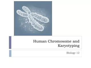
KARYOTYPING. A karyo type (Greek karyon = kernel, seed or nucleus) is the number and appearance of chromosomes in the nucleus of a eukaryotic cell. OR complete set of chromosomes in a species, or an individual organism. part of cytogenetics. Length, the position of the centromeres,

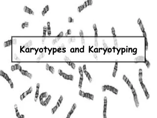
KARYOTYPING
E N D
Presentation Transcript
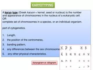
KARYOTYPING A karyotype (Greek karyon = kernel, seed or nucleus) is the number and appearance of chromosomes in the nucleus of a eukaryotic cell. OR complete set of chromosomes in a species, or an individual organism. • part of cytogenetics. • Length, • the position of the centromeres, • banding pattern, • any differences between the sex chromosomes, • any other physical characteristics. karyogram or idiogram
The study of karyotypes is important for • cell biology • genetics, • Clinical genetics • evolutionary biology • To study chromosomalaberrations, cellular function, taxonomic relationships, • and to gather information about past evolutionary events. • The sex of an unborn fetus can be determined
position of centromere - arm length ratio secondary constrictions (nucleolarorganisers) Short arm is labeled P (French for petit) Long arm is labeled Q
Human chromosomes are divided into 7 groups & sex chromosomes A 1-3 Large metacentric 1,2 or submetacentric B 4,5 Large submetacentric, all similar C 6-12, X Medium sized, submetacentric - difficult D 13-15 medium-sized acrocentric plus satellites E 16-18 short metacentric 16 or submetacentric 17,18 F 19-20 Short metacentrics G 21,22,Y Short acrocentrics with satellites. Y no satellites.
A B
STAINING The study of karyotypes is made possible by • cells (WBCs) arrested during cell division by a solution of colchicine. • Staining, Usually, a suitable dye, such as Giemsa • Colchicine inhibits microtubule polymerization by binding to tubulin, one of the main constituents of microtubules. Availability of tubulin is essential to mitosis, and therefore colchicine effectively functions as a "mitotic poison" or spindle poison. • For humans, white blood cells are used most frequently because they are easily induced to divide and grow in tissue culture. Sometimes observations may be made on non-dividing (interphase) cells.
Observations • 6 different characteristics of karyotypes are usually observed and compared • Differences in absolute sizes of chromosomes. • Chromosomes can vary in absolute size by as much as twenty-foldbetween genera of the same family. • 2. Differences in the position of centromeres. These differences probably came about through translocations. • 3. Differences in relative size of chromosomes. These differences probably arose from segmental interchange of unequal lengths.
Differences in basic number of chromosomes. • these differences could have resulted from successive unequal translocations which removed all the essential genetic material from a chromosome, permitting its loss without penalty to the organism (the dislocation hypothesis) or through fusion. Humans have one pair fewer chromosomes than the great apes. Human chromosome 2 appears to have resulted from the fusion of two ancestral chromosomes, and many of the genes of those two original chromosomes have been translocated to other chromosomes.
Differences in number and position of satellites. • Satellites are small bodies attached to a chromosome by a thin thread • Differences in degree and distribution of heterochromatic regions. • Heterochromatin stains darker than euchromatin. Heterochromatin is packed tighter. Heterochromatin consists mainly of genetically inactive and repetitive DNA sequences. Euchromatin is usually under active transcription.
Fundamental number • The fundamental number, FN, of a karyotype is the number of visible major chromosomal arms per set of chromosomes. Thus, FN ≤ 2 x 2n, why??? • Humans have FN ??? • (acrocentric chromosome pairs: 13, 14, 15, 21 and 22)
CHR NO Changes during development Instead of the usual gene repression, some organisms go in for large-scale elimination of heterochromatin, or other kinds of visible adjustment to the karyotype. • Chromosome elimination. In some species, as in many sciarid flies, entire chromosomes are eliminated during development. • Chromatin diminution . In this process, found in some roundworms, portions of the chromosomes are cast away in particular cells. This process is a carefully organised genome rearrangement where new telomeres are constructed and certain heterochromatin regions are lost. In A. suum, all the somatic cell precursors undergo chromatin diminution.
X-inactivation. The inactivation of one X chromosome takes place during the early development of mammals • In placental mammals, the inactivation is random as between the two Xs; thus the mammalian female is a mosaic in respect of her X chromosomes. In marsupials it is always the paternal X which is inactivated. In human females some 15% of somatic cells escape inactivation.
Ploidy: number of complete sets of chromosomes in a cell. • Polyploidy, more than two sets of homologous chromosomes in the cells, occurs mainly in plants. major significance in plant evolution • Haplo-diploidy: where one sex is diploid, and the other haploid. in the Hymenoptera. • Endopolyploidy occurs when in adult differentiated tissues the cells have ceased to divide by mitosis, but the nuclei contain more than the original somatic number of chromosomes. e.g the endocycle (endomitosis or endoreduplication) chromosomes in a 'resting' nucleus undergo reduplication, the daughter chromosomes separating from each other inside an intact nuclear membrane. • Aneuploidy:Aneuploidy is the condition in which the chromosome number in the cells is not the typical number for the species
classic" karyotype, CHROMOSOMAL STAINING • G-banding is obtained with Giemsa stain following digestion of chromosomes with trypsin. It yields a series of lightly and darkly stained bands — • the dark regions tend to be heterochromatic, late-replicating and AT rich. • The light regions tend to be euchromatic, early-replicating and GC rich. • This method will normally produce 300–400 bands in a normal, human genome. • R-banding is the reverse of G-banding (the R stands for "reverse"). The dark regions are euchromatic (GC rich regions) and the bright regions are heterochromatic (AT rich regions).
C-banding: stains centromeres. • Q-banding is a fluorescent pattern obtained using quinacrine for staining. The pattern of bands is very similar to that seen in G-banding. • T-banding: visualize telomeres. • Silver staining: Silver nitrate stains the nucleolar organization region-associated protein. This yields a dark region where the silver is deposited, denoting the activity of rRNA genes within the NOR.
SPECTRAL KARYOTYPING molecular cytogenetic technique used to simultaneously visualize all the pairs of chromosomes in an organism in different colors. • Fluorescently labeled probes • Mixtures
DIGITAL KARYOTYPING/ VIRTUAL KARYOTYPING Digital karyotyping is a technique used to quantify the DNA copy number on a genomic scale. Short sequences of DNA from specific loci all over the genome are isolated and typed.
RESTRICTION MAPPING • the process of obtaining structural information on a piece of DNA by the use of restriction enzymes. A restriction map is a map of known restriction sites within a sequence of DNA Restriction mapping steps Breaking DNA into pieces identifying the locations of the breakpoints.
Restriction Enzymes • endonucleases that recognize specific 4 to 8 base regions of DNA. restriction sites. • evolved as a bacterial defense against DNA bacteriophage • Recognizes Palindromicseq, • each strand of the DNA can self-anneal and the DNA forms a small cruciform structure • Hundreds of restriction enzymes that have been isolated and each one recognizes its own specific nucleotide sequence.
Sites for each restriction enzyme are distributed randomly throughout a particular DNA stretch. Digestion of DNA by restriction enzymes is very reproducible; every time a specific piece of DNA is cut by a specific enzyme, the same pattern of digestion will occur.
Uses of Restriction Mapping for many techniques used to manipulate DNA. One application is to cut a large piece of DNA into smaller fragments to allow it to be sequenced. Genes and cDNAs can be thousands of kilobases long (megabases - Mb); however, they can only be sequenced 400 bases at a time. DNA must be chopped up into smaller pieces and sub cloned to perform the sequencing.
Also, restriction mapping is an easy way to compare DNA fragments without having any information of their nucleotide sequence.
The sum of the individual fragments =size of the original fragment • If not there are two likely problems. • In one case, some of the smaller fragments may have run off the end of the gel. • the gel was not dense enough and therefore was unable to resolve fragments close in size. • This leads to a lack of separation of fragments which were close in size.