Download
1 / 20
260 likes | 1.33k Views
Lumbar Spinal Stenosis. Diagnosis and Management: A 30 year legend. Philip R. Weinstein, MD UCSF Dept. of Neurosurgery San Francisco, California. My Legends. Brain WR, Wilkinson M, eds. Cervical Spondylosis and other Disorders of the Cervical Spine, Phila. Saunders, 1967

E N D
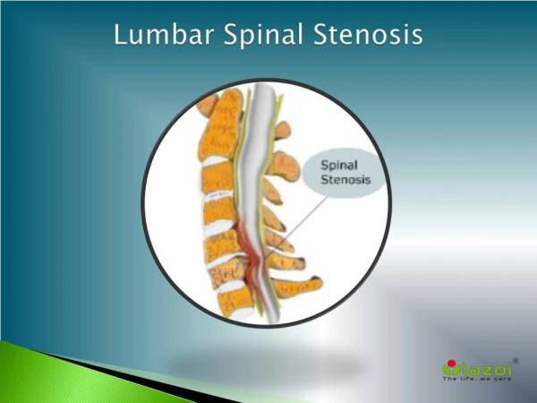
Lumbar Spinal Stenosis Diagnosis and Management: A 30 year legend Philip R. Weinstein, MD UCSF Dept. of Neurosurgery San Francisco, California
My Legends • Brain WR, Wilkinson M, eds. Cervical Spondylosis and other Disorders of the Cervical Spine, Phila. Saunders, 1967 • Wilson, CB: Significance of the small Lumbar Spinal Canal: Cauda Equina Compression Syndromes due to Spondylosis. III. Intermittent Claudication J Neurosurg 31 :499, 1969
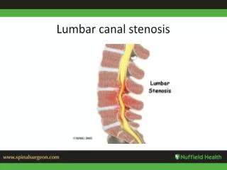
Classic case: 74 yo male; postural + ambulation claudication, bilat. footdrop, calf/thigh atrophy, incontinence
Why so mysterious? • Lumbar spondylostenosis, one of the most common neuro-spinal disorders, was not regularly recognized and treated until 1960. The diagnosis was not considered as an alternative to disc, infection or tumor because: • Stenosis not described/identified • Spondylosis not appreciated/visualized • Postural radiculopathy not understood • Atypical leg pain not interpreted • Neurogenic claudication not defined/explained • Cauda equina syndrome not diagnosed • Role of associated LBP/deformity not appreciated
Why has treatment been unsuccessful (20-40%)? • Clinical history/diagnosis not appreciated • Anatomy/imaging misinterpreted • Surgical plan incomplete or excessive • Surgical decompression inadequate • Fusion/fixation omitted/incomplete • Post-operative instability; ASD • Neuropathic radiculopathy • Progressive disc/facet DJD, spondy, scolio • Recurrent/progressive stenosis • Co-morbidities
Books on Lumbar Stenosis • Neurogenic Intermittent Claudication, Verbiest H, 1976, Elsevier • Lumbar Spondylosis, Weinstein P, et al, 1977, Year Book Medical • Cheirolumbar Dysostosis, Wachenheim A, et al, 1980, Springer • Lumbar Spinal Stenosis, Postacchini F, et al, 1989, Springer • Lumbar Spinal Stenosis, Andersson GBJ, et al, 1992, Mosby-Year Book • Lumbar Spinal Stenosis, Gunzberg R, et al, 2000, Lippincott-Williams&Wilkins
Evolution of a Concept: 1911-’25-’60Spondylotic Caudal Radiculopathy (SCR) • Bailey P, Casamajor L, Osteoarthritis of the Spine as a Cause of Compression of the Spinal cord and its roots: With report of five cases, J. Nerv. and Mental Dis. 38:588, 1911 • Parker JL, Adson AW, Compression of the Spinal Cord and its roots and hypertrophic Osteoarthritis, Surg. Gynec. Obstet. 41:1, 1925
Pioneering Intra-operative Measurements: Define the Syndrome Confirm the Diagnosis • Verbiest H, Further experiences on the Pathological influence of a Developmental narrowness of the bony Lumbar Vertebral canal, J. Bone Joint Surg. 37B:576, 1955
Developmental Variations/Stenosis Documentedin Normal Sized Cadavers • Epstein BS, Epstein JA, Lavine L, The Effect of Anatomic variations of the Lumbar vertebra and Spinal canal on Cauda equina nerve root syndromes, Am. J. Roentgenol. Radium Ther. Nucl. Med. 91:105, 1964
Vertebral Embryology • Unique paired growth centers for neural arch; vertebral body centers unite at 9 weeks • Premature arrest only dorsally in LS; both arch and body in dwarfism • Conus reaches L2; 22 weeks gestation • Most rapid canal growth; 18-36 weeks • L3-4 canal 80% at birth; 100%-1year; stops • L5 canal 50% at birth; 100%-5years • Canal deficiency: birth age/weight, mater. age • L5 trefoil canal: 15-25% Papp T, et al, JBJS 1995, 77B:469-472 Angevine JB, Clin. NS 1973, 20:95-113
Familial Cases Developmental non-dwarf(2 brothers and a sister) Postacchini F, et al, JBJS 67A:321, 1985
Incidence of Lumbar Stenosis in radiculopathy cases • Primary stenosis • 2% • Primary disc • 31% • Primary degenerative • 28% • Combined • 39% N=227 Paine K, Haung P, Lumbar Disc Syndrome JNS 37:75, 1972
Imaging Pearls for the Diagnostic Necklace • Thin section axials; no skipped levels • Compare disc vs mid-body sections • View images; beware report omissions/LRS • Sagittals for foraminal height/lat. osteophytes • Sagittals for disc height, spondylolisthesis • Coronals for scoliosis/foramen stenosis • Add CT to MRI for bone detail/facet tropism • CT myelo for postop cases/instrumented fusions • 3D CT to rule out pseudarthrosis • IV contrast CT for foraminal root constriction Radiculogram for foraminal fibrosis (TFESI) • Flexion-extension MRI for borderline cases • Intraop fluoro-CT useful for complex stenosis/deformity
Quantification of stenosis correlates with symptoms • Mean L4-5 canal area by CTM in extension in normals = 145mm2 (range 86-230) • Myelographic block occurs = 40mm2 • Transverse canal diameter below 11mm is symptomatic • Lateral recess height 2-4mm is symptomatic Wilmink JT, AJNR, 1989,26:173-181 Wilmink JT et al, Neuroradiology, 1988,3:5476-550
Radiographic-Clinical Correlation: Limitations of Measurements • Asymptomatic abnormalities seen • Magnification is variable • Imaging window, slice thickness, scan angle, alter bone/soft tissue measurements • Flex-ext. changes relationships • Ca on MR; CSF on CT not well visualized
Mechanisms of neurogenic claudication: compression, ischemia or both • 83yo male with L4-5 spondy II and stenosis 5yr. hx of leg pain during 1block walk relieved by standing • At autopsy: radicular arteries straightened veins compressed, neuronal loss, empty axons, demyelinization, arachnoid fibrosis, adjacent AV coiling-anastomosis Watanabe R, Parke W, JNS 64:64-70, 1986
Problems in Patient Selection and Surgical Planning • Patient age/co-morbidities • Previous surgery • Unilateral or bilateral decompression • “Asymptomatic” levels • MIS vs open • Disc “herniation” • Disc collapse/foramen stenosis • Spondylolisthesis/scoliosis/kyphosis • “Back pain” without instability (arthrogenic vs radicular) • Fusion: instrumented/PSF vs interbody fusion
What do Evidence-based Guidelines Tell Us? • Surgery resulted in better improvement in pain and function than non-operative rx for stenosis/deg. spondy @ 2yrs. 17% crossover to surg. Weinstein JN, (SPORT STUDY) NEJM, 2007;356(22):2257-70 • Surgery better for leg pain and back related function but equal to non-op rx for pt. satisfaction, back pain and primary sx relief @8-10yrs. 39% non-ops had surg. 23% reops. Atlas SJ, (MAINE STUDY) Spine, 2005;30(8):936-43
Clinical Trial Results • Unilat Laminotomy for Bilateral Microdecompr: 520 levels/374pts • 88% improved VAS/Prolo scale 5yr f/u 0.8% instability: none reoperated Costa F etal. JNS-Spine 2007;7(6):579-586