Download

1 / 13
150 likes | 589 Views
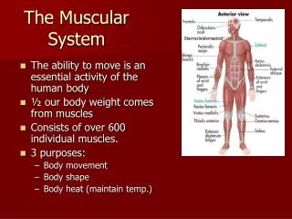
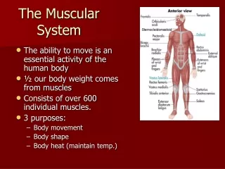
The Muscular System. Approximately 40% of your body weight is your muscle. Functions Muscles produce movement. When muscle contracts , it pulls insertion bone near origin bone. Movement occurs at joint between origin and insertion .

E N D
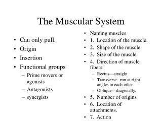
The Muscular System Approximately 40% of your body weight is your muscle. Functions Muscles produce movement. When muscle contracts , it pulls insertion bone near origin bone. Movement occurs at joint between origin and insertion. Origin– The bone that moves less, provides the area of attachment for the end of the muscle called the origin.
Insertion – the movable bone provides the surface for the muscle’s insertion. e.g. biceps : origin at the joint of humerus and scapula, insert on radius. triceps : origin at humerus, scapula and clavicle, inserts on ulna. • Biceps and triceps work in opposing pairs in an antagonistic system. • Groups of muscles usually contract to produce a single movement – synergistic pattern. e.g. extension of lower legs is by rectus femoris, gracilis and sartorius.
Reference: http://www.ultranet.com/~jkimball/BiologyPages/M/Muscles.html#Anatomy_of_Skeletal_Muscle http://www.lrn.org/Content/Lessons/muscle.html#overview
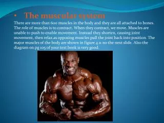
There are three different types of muscles a. skeletal muscle – voluntary, striated muscle. b. cardiac muscle – involuntary, striated (branching) muscle. c. smooth muscle – involuntary, non-striated muscle, e.g. Stomach, intestinal tract, urinary bladder and blood vessels. Reference: http://www.terrebonnehigh.com/science/biol2n9.htm
Skeletal Muscle Contraction Skeletal muscles contain thousands of muscle fibers (muscle cells). Each fiber consists of finer threadlike structures called myofibrils. Myofibrils contain two kinds of protein strands: thick filament, myosin, with side projecting cross-bridge. Thinner filament, actin. Repeating bands of actin and myosin translate into light – dark repeating unit that gives skeletal muscle its striped appearance. Dark line (Z) line between each repeating unit is defined as sacromere that is the fundamental unit of muscle contraction.
References: http://www.uoguelph.ca/zoology/devobio/210labs/sketchmuscle1.html http://www.lrn.org/Content/Lessons/muscle.html#overview
Muscle Structure Reference: http://members.shaw.ca/bodybuilding/Muscles/structure.html
Reference: http://www.ultranet.com/~jkimball/BiologyPages/M/Muscles.html#Anatomy_of_Skeletal_Muscle http://www.lrn.org/Content/Lessons/muscle.html#overview
The Sliding-Filament Model Reference: http://www.ultranet.com/~jkimball/BiologyPages/M/Muscles.html#Anatomy_of_Skeletal_Muscle
Role of Calcium in Muscle Excitation and Contraction Coupling http://cwx.prenhall.com/bookbind/pubbooks/martinidemo/chapter10/medialib/CH10/html/ch10_4_1.html
Role of Calcium in Muscle Excitation and Contraction Nerve ending release neuro transmitter at neuro-muscular junction – membrane excitation Sarcoplasmic reticulum release Ca 2+ Ca 2+ binds to troponin removing blocking action of tropomyosin Actin and myosin –cross bridge movement ATP is required Remove Ca 2+ from troponin restores blocking action of tropomyosin Ca2+ uptake (ATP required)
Muscle Disorders • Muscular dystrophy – progressive weaken of the muscles. • Paralysis – loss of ability to produce voluntary movement. This is due to disease or injury of brain or spinal cord or nerve • Muscle atrophy – muscle shrinkage. Decrease in muscle size. • Muscle hypertrophy – increase in muscle size because of over work. e.g. heart frequently hypertrophy from over work.
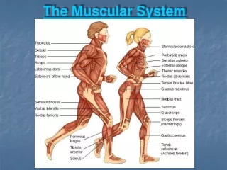
Muscle StructureFront View Back View Reference:http://www.rrcc.cccoes.edu/academic/health/fitnesscenter/muscle.htm