MOTOR CORTEX
340 likes | 1.3k Views
Motor cortex, descending tracts (Pyramidal and Extrapyramidal tract)

MOTOR CORTEX
E N D
Presentation Transcript
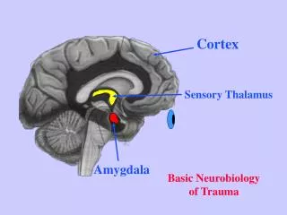
Motor cortex and Descending pathways D.A. Asir John Samuel,BSc (Psy), MPT (Neuro Paed), MAc, DYScEd, C/BLS, FAGE Lecturer, Alva’s college of Physiotherapy, Moodbidri
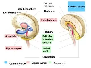
Lobes of cerebral cortex • Frontal lobe – in front of central sulcus or fissure of Roland • Parietal lobe – central sulcus/parieto-occipital sulcus • Occipital lobe – behind parieto-occipital cortex • Temporal lobe – below lateral or sylvian fissure
Lobes of cerebral cortex • Frontal lobe – in front of central sulcus or fissure of Roland • Parietal lobe – central sulcus/parieto-occipital sulcus • Occipital lobe – behind parieto-occipital cortex • Temporal lobe – below lateral or sylvian fissure
Frontal lobe • Broadmann’s Area 4 – precentral gyrus and is Primary motor Area • Supplementary motor Area – ant. to Areas 4 & 6 • Premotor Area (Area 6) – ant. to Area 4 • Broca’s Area (Area 44) – motor area for speech, in post. part of frontal operculum in domi. H • Frontal eye field (Area 8)
Broadmann’s Area 4 • Primary motor area • Occupies posterior part of precentral gyrus and ant.lip and walls of central sulcus • Highest centre for voluntary movements • Gives origin to fibres of pyramidal tract and other descending tract • Interconnected to Area 6 and sensory area
Broadmann’s Area representation • Size of cortical area is proportional to functional importance and activity of region • Order of representation from medial to lateral surface is toes, ankle, knee, hip, trunk, shoulder, arm, elbow, wrist, hand, fingers, thumb, eyes, face, jaw and tongue • Motor homonuclus
Supplementary motor area • Medial surface of hemisphere in front of area 4 and 6, in medial frontal gyrus • Representation is head to foot anteroposterly • Provides background for fine complex movements from motor cortex
Area 6. Premotor area • Anterior to area 4 and in medial aspect of H • Planning and rehearsal takes place before execution • Gives fibres to pyramidal tract and basal ganglia concerned with postural control • Postural background for complex coordinated movements
Frontal eye field (Area 8) • Middle frontal gyrus • Receives association fibres from occipital cortex (Areas 18, 19) • Projects of oculomotor nuclei • Moving head and eyes towards the objects to be seen
Layers of cerebral cortex • Molecular layer (Plexiform layer) • External granular layer • External pyramidal layer • Internal granular layer • Ganglionic layer (Internal pyramidal layer) • Multiform layer (layer of polymorphic cells)
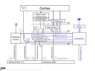
Descending pathways • Corticospinal (Pyramidal) and corticobulbar tracts • Vestibulospinal • Reticulospinal • Tectospinal • Rubrospinal • Olivospinal
Corticospinal tract • Arise from axons of pyramidal cells situated in 5th layer of cerebral cortex • Primary motor area – 30% • Premotor area – 30% • Postcentral gyrus (Parietal lobe) – 40%
Corticospinal tract Descend in corona radiata and converge in posterior limb of internal capsule In midbrain, occupies middle 3/5 of basis pedunculi (crus cerebri) In pons, broken up into small no.of bundles
Corticospinal tract In lower part of Pons, fibres reunite to form compact bundle Descends in ventral part of medulla forms Prominent Pyramids of Medulla
Corticospinal tract At junction of MO & SC, most fibres cross midline at decussation of Pyramids Form Lateral corticalspinal tract Uncrossed – Anterior corticospinal tract Terminates in anterior gray column, by internuncial
Corticospinal tract • Speed and agility to voluntary movements • Rapid skilled movements • Upper motor neuron lesion • Spasticty
Vestibulospinal tract • Vestibular nuclei are situated in pons and MO beneath floor of 4th ventricle` • Neurons of lateral vestibular nucleus give rise to axons that form VST • Tract descends uncrossed though medulla and through length of SC in ant.white column
Vestibulospinal tract • Ventral or anterior VST • Lateral VST • Regulate muscle tone and equilibrium • Facilitates activity of extensor muscles and inhibit activity of flexor muscles
Reticulospinal tract • Throughout midbrain, pons and MO, groups of scattered nerve cells and nerve fibers exist that are collectively known as reticular formation • Pontine reticulospinal tract • Medullary reticulospinal tract
Reticulospinal tract • From pons, neurons send axons, which are mostly uncrossed, down into SC and form pontinereticulospinal tract (medial RST) • From medulla, which are crossed and uncrossed, to SC and form medullary reticulospinal tract (lateral RST)
Reticulospinal tract • Reticulospinal fibres from pons descend through ant.white column • While from MO descend in lateral white column • Both sets enter AGC and may facilitate or inhibit activity of α and β
Reticulospinal tract • Influence voluntary movements and reflex activity • Provide a pathway by which hypothalamus can control sympathetic outflow and sacral parasympathetic outflow
Rubrospinal tract • Arises from posterior 1/3rd or nucleousmagnocellularis of red nucleus in midbrain • On leaving RN, fibres cross to opposite side in tegmentum of midbrain as Forel’sdecussation • Descend through reticular formation of pons and medulla to SC
Rubrospinal tract • Lies anterior to lateral corticospinal tract • Prominent upto mid thoracic region • Receives impulses from cerebral cortex, cerebellum and corpus striatum • Part of lateral motor system • Facilitates flexor muscle and inhibits ext./AG
Tectospinal tract • Arise in superior colliculus of midbrain • Cross over to opposite side in tegmentum as Meynert’sdecussation • Descend through RF of pons and medulla
Tectospinal tract • Superior colliculus receives fibres from retina • Coordinate retinal impulses with body movements and reflex postural movements
Olivospinal tract • Arise in inferior olivary nucleus of medulla • Descend in anterolateral part of white column • Traced only as far as cervical region