Download
1 / 48
490 likes | 756 Views
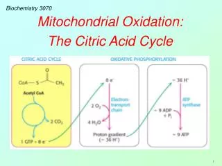
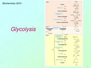
Biochemistry 3070. Oxidative Phosphorylation. Oxidative Phosphorylation. Glycolysis and the citric acid cycle yield NADH and FADH 2 . Both these electron carriers are energy-rich molecules because their electrons have a high transfer [redox] potentials.

E N D
Biochemistry 3070 Oxidative Phosphorylation
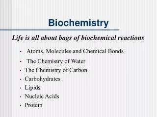
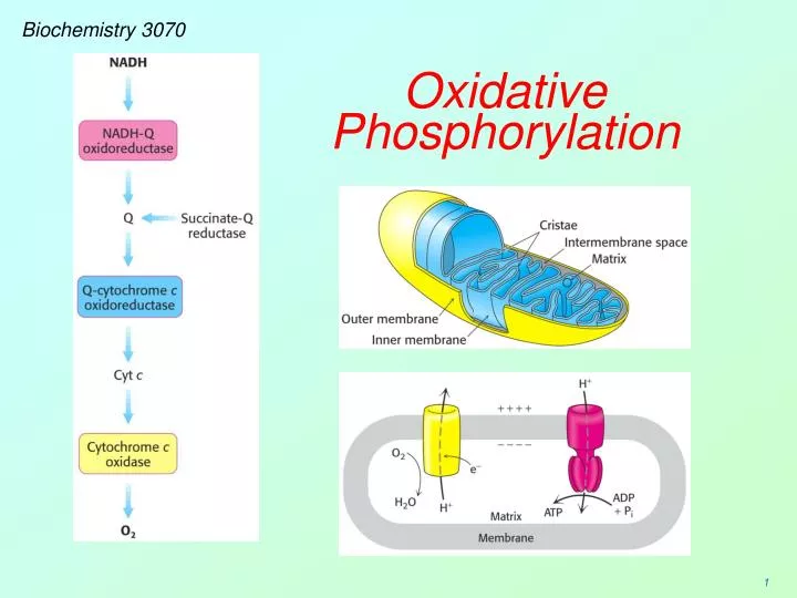
Oxidative Phosphorylation • Glycolysis and the citric acid cycle yield NADH and FADH2. • Both these electron carriers are energy-rich molecules because their electrons have a high transfer [redox] potentials. • Oxidative phosphorylation is the process of converting this high redox potential into energy-rich ATP molecules. • This process, together with the reactions that form the electron carriers is often called respiration.
Oxidative Phosphorylation ATP synthesis via oxidative phosphorylation occurs via two separate systems in the mitochondrion. • Electrons are “transported” via numerous membrane-bound carriers from NADH to O2. During these reactions, a proton gradient is formed across the mitochondrial inner membrane. • The proton-motive force in the gradient is then harnessed to produce ATP.
Oxidative Phosphorylation • Mitochondrial structure plays a critical role in forming and utilizing the proton gradient to synthesize ATP. • Protons are “pumped” from the matrix across the inner membrane into the intermembrane space. • ATP is synthesized in the matrix, as protons flow back through the membrane.
Oxidative Phosphorylation • First, we will learn how electrons are transported from NADH/FADH2 to oxygen with the concomitant formation of the proton gradient. Then, we will see how ATP is made. • Around 1961, Peter Mitchell published a famous theory that ATP was synthesized by proton gradients formed in the mitochondrion: The “chemiosmotic hypothesis.” • Initially, Mitchell found little support for his proposal, but when no one could prove him wrong, more believers were “converted” and he eventually received the Nobel Prize for his work in 1978. http://nobelprize.org/chemistry/laureates/1978/
Oxidative Phosphorylation The driving force for electron transfer from NADH to O2 is explained by their reduction potentials. Recall from freshman chemistry that the reduction potential of redox-active substances is easily measured with a volt meter and two separate chambers. Published reduction potentials for NADH and O2 show that electrons flow spontaneously from NADH to O2, with a combined redox potential of +1.14 Volts: ½ O2 + 2 H+ + 2e- → H2O E’0 = +0.82 V NAD+ + H+ + 2e- → NADH E’0 = - 0.32 V ½ O2 + NADH → H2O + NAD+ E’0 = +1.14 V
Oxidative Phosphorylation Rather than occurring in a single step, electrons from NADH pass through groups of carriers, mostly within the mitochondrial inner membrane, eventually reaching oxygen. The most interesting of these carriers are three groups of protein complexes often identified as “Sites I, II, III, & IV.” I II III IV
Oxidative Phosphorylation Sites I, II, III, and IV each contain numerous protein subunits: I II III IV
Oxidative Phosphorylation Electrons from NADH pass through Site I: During their transit, electrons pass through a flavin [isoalloxazine] ring and iron-sulfur clusters. The reduction and subsequent oxidation of the isoalloxazine requires two hydrogen atoms. Due to the unique structure of the NADH-Q oxidoreductase complex, hydrogens are drawn from the matrix during reduction and then ejected into the intermembrance space during oxidation. Matrix:H+ H+ Intermembrane Space:H+ H+
Oxidative Phosphorylation Electrons are transported through the membrane from Site 1 to Site 3 by ubiquinol (ubiquinone). This fat-soluble carrier picks up electrons from Site 1 (Q→QH2), then diffuses laterally through the membrane delivering electrons to Site 3 (QH2→Q), then returns to Site 1 for more.
Oxidative Phosphorylation QH2 delivers protons to Site III: “Q-Cytochrome c Oxidoreductase:” This complex moves electrons through a variety of carriers, including 3 hemes and another 2Fe-2S cluster. As in Site 1, protons are taken from the matrix and deposited into the intermembrane space during passage of electrons through this site and reduction of the next carrier, cytochrome c:
Oxidative Phosphorylation • Cytochrome c (“cyt c”) is a water-soluble protein with a single heme prosthetic group. • Each cyt c transports a single electron from Site III to Site IV through the aqueous intermembrane space. (Cyt c is the only electron carrier that is not located in the mitochondrial membrane!) • Cyt c is structurally similar in many organisms, so much so, that cyt c from one organism can sometimes be used in vitro to transport electrons from Site III→IV in electron transport pathways of other organisms!
Site IV (“Cytochrome oxidase”) takes electrons from cytochrome c and uses them to reduce oxygen to water. As in previous sites, obligatory hydrogen withdrawal from the matrix accompanies this final electron transport process:
Oxidative Phosphorylation Additionally, extra protons are “pumped” from the matrix into the innermembrane space during the oxidation of cytochrome c.
Oxidative Phosphorylation This pathway of electron transport from NADH to H2O through a variety of electron carriers allows the transformation of redox energy into a proton gradient. How do scientists know the order of the electron carriers in this pathway? The reduction potential (E’0) of each carrier has been determined over the years. Once a reduction potential for a carrier is known, it is relatively easy to place it in its correct position relative to the others since electrons move spontaneously from carriers with lower reduction potentials to carriers with higher reduction potentials.
Oxidative Phosphorylation Note the different E’0 for NAD+ and FAD: FAD is not a strong enough reductant to reduce electron carriers at Site 1. However, it can reduce ubiqinone.
Oxidative Phosphorylation • FADH2 delivers electrons to the electron transport pathway by reducing ubiquinol (Q) to ubiquinone (QH2). • Recall that FADH2 is formed in the TCA cycle when succinate is oxidized to malate. Electrons from FADH2 reduce Q to QH2 and flow through electron transport, ending up on oxygen. • Consequently, less ATP is formed from the oxidation of FADH2 than from NADH. FADH2
Oxidative Phosphorylation – Electron Carrier Postioning in Chain The pathway of electron transport is easily determined by comparing reduction potentials (E’0):
Oxidative Phosphorylation – Electron Carrier Postioning in Chain Diagram of electron transport chain and electron flow. (Garrett & Grisham, Biochemistry, 3rd ed., Brooks/Cole) FADH2 FAD
Another View of Electron Transport (Mathews, et.al, Biochemistry, 3rd ed., Addison Wesley)
Oxidative Phosphorylation How is the proton-motive force created by obligatory proton transport during passage of electrons converted into high-energy phosphate bonds in ATP? The historical aspects of the discovery of this fascinating process deserves our attention.
Oxidative Phosphorylation Ephriam Racker (Cornell) discovered unique knob-like structures on the matrix side of the inner membrane. He removed these knobs with mild detergents and mixed them with ATP. The ATP was immediately hydrolyzed to ADP, so he named the knobs “ATPase.”
Oxidative Phosphorylation Protons flow through the channel (F0) to the large knob (F1)where ATP is synthesized. The “ATPase” knobs described by Racker destroy ATP by converting it into ADP + Pi if they are not attached to the F0 subunit. When attached, they catalyzed the opposite reaction, namely ATP synthesis.
Oxidative Phosphorylation In Ephriam Racker’s personal account of his discovery, he recites that once removed, the ATPase knobs were difficult to reconsititute, because they were working in the cold room at ~ 4°C. The ATP knobs are labile at this temperature. A new student tried a serendipitous experiment at room temperature and was able to reattach the knobs, restoring normal ATP synthesis.
Oxidative Phosphorylation Racker always sought to encourage young students and offered some encouraging advice (“lessons” he’d learned). Examples from a lecture series at Pomona College in 1973: • Chemistry is good, nature is better. • A clean experiment is worth more than a few hundred dirty calculations. • It doesn’t matter if you fall down as long as you pick up something from the floor while you get up. • Not everything that shines is gold. Not everything that floats after high-speed centrifugation is soluble. • Progress is made by young scientists who carry out experiments old scientists said wouldn’t work. • If you accept the statement that only uninhibited investigators use inhibitors, you will soon find out what kind of people work in the field of oxidative phosphorylation. • “I have yet to see a problem however complicated that, when you look at it the right way, does not become more complicated.” (Paul Aldeston)
Oxidative Phosphorylation • Racker was sure ATP was synthesized by proton flow through a membrane channel that was linked to the “ATPase knobs.” • Other scientists were skeptical. • Racker joined with Walt Stoeckenius (UCSF) to conduct a brilliant experiment. • Walt was having a difficult time convincing people that that “red-tide” bacteria Halobacterium halobium was using light to drive a proton pump via a membrane protein (bacteriorhodopsin).
Proton Motive Force & ATP Synthesis • They formed artifical bilayer lipid membrane vesicles and inserted both bacteriorhodopsin and the F0 & F1 coupling factors from mitochondria. • When they turned on the light, ADP was converted into ATP! • Both scientists proved their hypotheses in this elegant experiment.
Oxidative Phosphorylation – Proton Flow In mitochondria, protons flow from the intermembrane space back into the matrix via a specialized channel and the ATPase “knobs,” where ATP is synthesized.
Oxidative Phosphorylation – Proton Flow (Garrett & Grisham, Biochemistry, 3rd ed., Brooks/Cole)
ATP synthesis • The addition of phosphate to ADP seems simple: • Yet, the mechanism and the protein actions are quite complex. • Parts of the ATP synthase complex actually rotate, forming ATP as they turn.
ATP Synthase Structure & Function The 9-12 cylinder-shaped proteins forming the c-ring are imbedded deep in the membrane. Each of these protein subunits has a critically important amino acid residue [asp 61] about half way across the membrane that is required for the system to function. Subunit a is where protons pass through the complex from the intermembrane space into the matrix. Protons can not pass through unless they go through both channels.
ATP Synthase Structure & Function • Aspartic acid accommodates this proton transfer from one channel to the other. • An asp-61 in one c-ring subunt binds to the proton and then rotates around (the long way) where it is delivered to the other channel:
ATP Synthase Structure & Function The γ-subunit is tightly attached to the c-ring proteins. As such, it rotates with the c-ring during proton translocation. However, the three αβ pairs are held in place by the b2 arm. Therefore, the γ-subunit rotates inside the core of the F1 complex. As the γ-subunit rotates, it changes the conformation of the β subunits. This is an important facet of ATP synthesis.
ATP Synthase Structure & Function • A team of researchers led by John Walker crystallized the F1 (knob) structures, in the presence of ADP and App(NH)p (a non-hydrolyzable form of ATP.) • X-ray crystallography showed that the three β subunits were each bound to different substrates: • One to App(NH)p (the ATP analog) • One to ADP • One to nothing at all (empty site)
ATP synthesis • Paul Boyer finally put the puzzle together by proposing that there must be three sites with different binding affinities for the substrate (ADP + Pi) and product (ATP). • In fact, the three β-subunits interact in such a way that when one assumes the β-empty form, its neighbor to one side must assume the β-ADP form, and the other neighbor the β-ATP form. • Thus, one complete rotation of the γ-subunit causes each β-subunit to cycle through all three of its possible confomations, and for each rotation, three ATPs are synthesized and release from the enzyme surface. • Boyer received the Nobel Prize for this work in 1997 (born and raised in Provo, Utah) http://nobelprize.org/chemistry/laureates/1997/boyer-autobio.html
ATP synthesis – byer’s “Binding-change Model” Stryer, et.al, Biochemistry, 5th ed. (Mathews, et.al, Biochemistry, 3rd ed., Addison Wesley) (Garrett & Grisham, Biochemistry, 3rd ed., Brooks/Cole)
ATP synthesis • ATP moves from the mitochondrial matrix to the cytosol via a specialized membrane transport protein, “ATP-ADP translocase.” • Translocase is tightly coupled to the exchange of ADP for ATP as ATP exits.
ATP synthesis • Some NADH molecules are reduced in the cytosol and must be transported into the mitochonria for electrons to enter the electron transport pathway. • Two different “shuttles” are commonly encountered: • Glycerol 3-phosphate shuttle (transfers electrons to FADH2 . • Malate-aspartate shuttle (transfers electrons to NADH)
ATP synthesis Glycerol 3-phosphate shuttle: (NADH 2e- FADH2)
ATP synthesis • Malate-aspartate shuttle: (NADH 2e- NADH)
ATP Accounting Assumptions for Mitochondrial Oxidation (with G-3-P Shuttle): - - - - - - - - - - - - - - - - - - - - ATP Yields (2 Sig.Figs): NADH……… 2.5 ATP Succinate… 1.5 ATP Total: 30 ATP - - - - - - - - - - - - - - - - - - - - ATP Yields (1 Sig.Fig.): NADH………… 3 ATP Succinate….… 2 ATP Total: 36 ATP - - - - - - - - - - - - - - - - - - - -
ATP Uncoupling • ATP synthesis can be “uncoupled,” if the proton gradient is prematurely dissipated or impeded. • Certain inhibitors of electron transport act at specific sites to stop electron flow. • Site I: amytal & rotenone • Site III: antimycin A • Site IV: CN-, N3-, CO
ATP Uncoupling Two other inhibitors stop the flow of protons back through ATP synthase: • Oligomycin & • dicyclohexylcarbodiimide (DCCD) • Questions: • What would happen to the rates of electron transport and ATP synthesis if oligomycin were added to tightly-coupled, functioning mitochondria? • In a similar but separate experiment, what would happen to both these rates if the F1 portion (knobs) of the ATP synthase were removed?
ATP Uncoupling • Other substances allow protons to diffuse back across the membrane without going through the ATP synthase complex. • The toxic agent 2,4-DNP dissipates the proton gradient, slowing or even stopping ATP synthesis. • This phenol can carry protons across the inner mitochondiral membrane.
ATP Uncoupling • Hibernating animals also uncouple ATP synthesis to generate heat (nonshivering thermogenesis). • In brown adipose tissue (which is very rich in mitochondria), uncoupling protein (UCP) or “thermogenin,” forms a pathway for the flow of protons back into the matrix. • This short circuits the proton gradient, generating heat. • Some flowers also generate heat this way to voltalize fragrances that attract insects to fertilize their flowers.
End of Lecture Slides for Oxidative Phosphorylation Credits: Many of the diagrams used in these slides were taken from Stryer, et.al, Biochemistry, 5th Ed., Freeman Press (in our course textbook) and from prior editions of this text.