Download
1 / 24
250 likes | 393 Views
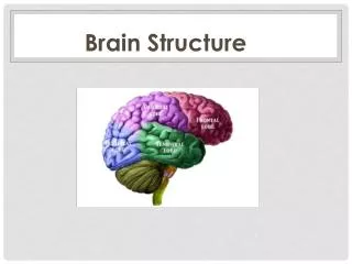
Quantitative Brain Structure Analysis on MR Images. Zuyao Shan, Ph.D. Division of Translational Imaging Research Department of Radiological Sciences. Outline. Introduction Cerebellum segmentation ( Preliminary study ) Cortical structure segmentation. Brain Segmentation.

E N D
Quantitative Brain Structure Analysis on MR Images Zuyao Shan, Ph.D. Division of Translational Imaging Research Department of Radiological Sciences
Outline • Introduction • Cerebellum segmentation (Preliminary study) • Cortical structure segmentation
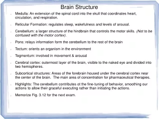
Brain Segmentation With the ability to identify brain structures on MR images and to detect anatomic changes, the new volumetric tools aid in the diagnosis, treatment, and elucidation of changes associated with disease or abnormality. • Registration – based approaches • Pros: Straightforward tenet, robustness • Cons: Accuracy limited by match quality, mismatch leading to significant errors, relying on image only. One-one mapping may not existed, Speed • Deformable model – based approaches • Pros: Prior knowledge incorporated, high accuracy. • Cons: Good initialization needed, identification of landmarks
Brain Segmentation: inter-personal variability • More challengesin pediatric patients with brain tumors: • Removal of tissues • Different stages of development • An adequate method should cope with high inter-subject variability with high accuracy
Brain Segmentation: Cerebellum Knowledge – guided active contour • Rigid-body registration: good initialization • Prior defined template: Knowledge incorporated • Active contour adjustment: high accuracy, robustness
Brain Segmentation: Cerebellum Active contour (Snake): energy-minimizing spline
Bending in the contour, low internal energy Small High Low Tension in the contour, low internal energy Small High Low Brain Segmentation: Cerebellum Active contour (Cont.): Internal energy
Brain Segmentation: Cerebellum Active contour (Cont.): External energy Distance Sobel edge detection transform
Brain Segmentation: Cerebellum Visual inspection
Brain Segmentation: Cerebellum Visual inspection
S1∩ S2 Brain Segmentation: Cerebellum • Similarity evaluation • Kappa index A vs. M1: ~ 0.94; A vs. M2: ~0.93; M1 vs. M2: 0.97 Compared with 0.77~0.84 for pediatric brain tumor patient in recent report1 • D’Haese P et al. Int J Radiat Oncol Biol Phys 2003; 57 (2 Suppl): S205
Brain Segmentation: Cortical Structures KAM, Knowledge-guided Active Model • New object functions In contrast, Registration – based approaches maximize S; deformable model – based approaches minimize H • Pediatric brain atlas • Affine registration (H) • 3D active mesh (S)
Brain Segmentation: Affine Registration 12 DOF: 3 translations, 3 rotations, 3 scaling, and 3 shearing
Brain Segmentation: Active Models External Energy: attract triangle vertex to the edge of the image
Brain Segmentation: Active Models Internal Energy: control the behavior of triangle mesh models
Brain Segmentation: Cortical Structures Segmentation results
Brain Segmentation: Cortical Structures Segmentation results
Brain Segmentation: Cortical Structures Segmentation results compared with SPM2 • Volumetric agreement: KAM : 95.4% ± 3.7% SPM2 : 90.4% ± 7.4% • Image similarities: KAM : 0.95 SPM2 : 0.86
Brain Segmentation: Summary • Pediatric brain atlas • www.stjude.org/brainatlas • KAM, Knowledge-guided Active Model • preliminary results indicate that when segmenting cortical structures, the KAM was in significantly better agreement with manually delineated structures than the nonlinear registration algorithm provided by SPM2.
Brain Segmentation: Future Studies • Validation of KAM • Application of KAM • Incorporating KAM into radiation therapy planning • Quantitative evaluation of cortical structure changes • Further development of KAM • Subcortical Structures • Brain Tumors
Acknowledgements Mentor: Dr. Wilburn E Reddick Colleagues: Dr. Robert J Ogg Dr. Fred H. Laningham Dr. Claudia M. Hillenbrand Carlos Parra, John Stagich, Dr. Qing Ji, John Glass, Jinesh Jain, Travis Miller, Rhonda Simmons