Download
1 / 12
130 likes | 142 Views
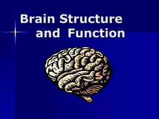
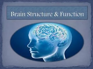
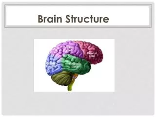
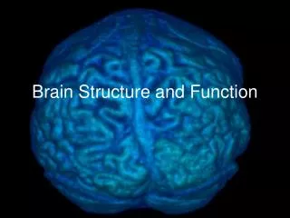
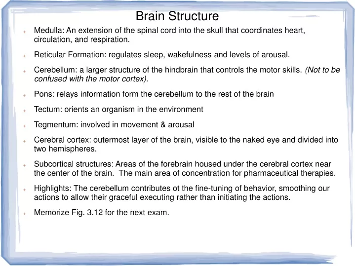
Brain Structure. Medulla: An extension of the spinal cord into the skull that coordinates heart, circulation, and respiration. Reticular Formation: regulates sleep, wakefulness and levels of arousal.

E N D
Brain Structure • Medulla: An extension of the spinal cord into the skull that coordinates heart, circulation, and respiration. • Reticular Formation: regulates sleep, wakefulness and levels of arousal. • Cerebellum: a larger structure of the hindbrain that controls the motor skills. (Not to be confused with the motor cortex). • Pons: relays information form the cerebellum to the rest of the brain • Tectum: orients an organism in the environment • Tegmentum: involved in movement & arousal • Cerebral cortex: outermost layer of the brain, visible to the naked eye and divided into two hemispheres. • Subcortical structures: Areas of the forebrain housed under the cerebral cortex near the center of the brain. The main area of concentration for pharmaceutical therapies. • Highlights: The cerebellum contributes ot the fine-tuning of behavior, smoothing our actions to allow their graceful executing rather than initiating the actions. • Memorize Fig. 3.12 for the next exam.
Subcortical Regions • Thalamus: relays and filters information from the senses and transmits the information to the cerebral cortex. All the sense except smell, which has direct connections to the cerebral cortex, indicating its evolutionary age. Also closes the pathways of incoming sensations during sleep. • Hypothalamus regulates body temperature, hunger, thirst, and sexual behaviour. Lesions here can lead to overeating or starvation. (Connect to WA2). • Pituitary gland: the master gland of the body's hormone-producing system, releasing hormones that direct the functions of many other glands in the body. • limbic system: composed of the hypothalamus, hippocampus & amydala; involved in motivation, emotion, learning & memory. • hippocampus: critical for creating new memories and integrating them into a network of knowledge so that they can be stored indefinitely in other parts of the cerebral cortex. • amygdala: a part of the limbic system that plays a central role in many emotional processes particularly the formation of emotional memories; the 'fear center' of the brain. • basal ganglia: a set of subcortical structures that direct intentional movements. • corpus callosum: a thick band of nerve fibers that connects large areas of the cerebral cortex; supports communication of information across the hemispheres.
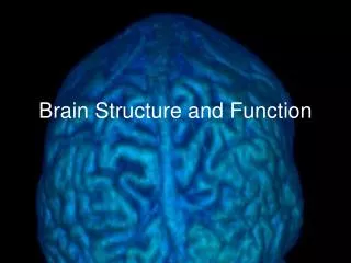
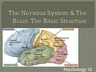
Cerebral Cortex • Memorize Fig. 3.14 cerebral cortex and lobes for the next exam. • The functions of the cerebral cortex can be understood at three levels: the seperation of the cortex into two hemispheres; the functions of each hemisphere; the role of the cortical areas. • Each hemisphere controls the functions of the opposite side of the body, or contralateral control. • The largest of the commissures is the corpus callosum, which allows information received in the right hemisphere to pass across and be registered (virtually instantaneously) in the left hemisphere. • the occipital lobe processes visual information. Sensory receptors in the eyes send information to the thalamus, which in turn sends it to the primary areas of the occipital lobe. When you hear of Neanderthals having larger brains than we do, it is this lobe. • the parietal lobe carries out functions that include processing information about touch. • the somatosensory cortex represents the skin areas of the contralateral surface of the body. • Memorize Fig. 3.16 ( somatosensory and motor cortices) for the next exam. • temporal lobe: responsible for language • frontal lobe and association areas: thinking, planning, memory & judgment.
Association Areas • Association areas: composed of neurons that help provide sense and meaning to information registered in the cortex. • Neurons in the primary visual cortex are highly specialized: horizontal orientation, movement, human vs non-human forms. • Neurons in the primary auditory cortex register sound frequencies, but areas in the temporal lobe allow those sounds to be given meaning. • Association areas weave together the threads of information in various parts of the cortex into a meaningful perception of the world. • Mirror neurons are active when an animal performs a motor behavior, and also when the animal observes another animal of its own species performing the same behavior. • Mirror neurons aid in recognizing the goal of that behavior and the outcome of that action. • The brain is plastic: functions that were assigned to certain areas of the brain may be capable of being reassigned to other (near) areas of the brain. Remember the homunculus as to how this works. • Extraordinary amounts of stimulus to a certain cortical area will re-organize it; there is greater plasticity within the motor cortex of professional musicians compared with non-musicians. • Physical exercise can increase the number of synapses, promoting development of new neurons in the hippocampus.
Phantom Limb Pain • Phantom Limb Pain: a syndrom where a patient can feel their limbs moving, even in co-ordinated gestures. Worse, some feel chronic cramps in missing limbs. • Researchers stimulated the skin surface in various regions around the face, torso, and arms while monitoring brain activity in amputees and non-amputees. • Brain imaging displayed the somatosensory cortex activated. Areas of the face and upper arm activated an area in the somatosensory cortex once activated by the missing hand. • Plasticity: the cortical representations for the face and the upper arm normally lie on either side of the representation for the hand. New contiguous mappings happened. • Some were quite precise: in some amputees, when specific areas of the facial skin were activated (eg: upper lip) the patient reported sensations in just one finger of the phantom hand. • Mirror box: teaches amputees to increase voluntary control over their phantoms limbs (necessary for the cramping problem). • The phantom hand appears to respond to motor commands; with practice the patient can become better at 'moving' the phantom in response to voluntary commands. • Haiti earthquake victims in 2012 were greatly helped by this simple technique.
Investigating the Brain • Much research in neuroscience correlates the loss of specific perceptual, motor, emotional & cognitive functions with specific areas of brain damage. • Broca described a patient who had lost the capacity to produce spoken language, but not the ability to understand language, due to damage in a small area in the left frontal lobe. • Wernicke described a patient with an impairment in language comprehension,but not the ability to produce speech, associated with damage to an area in the left temporal lobe. • Memorize 'the strange case of Phineas Gage' for the next exam. • Disorders can threaten the ability of the brain to function, such as severe, intractable epilepsy. • Seizures that begin in one hemisphere cross the corpus callosum to the opposite hemisphere and start a feedback loop that results in a 'firestorm' in the brain. • Split-brain procedure severes the corpus callosum = lateralized perception. • Memorize Sperry's split-brain experiment for the next exam. Note: the figure is a snapshot of less than a second's time. • Also important: each optic nerve has a left and right visual field (contrast this to the wrong idea that the left only gets info from the right).
Investigating the Brain • Chimeric Faces: When a person with a split brain views a chimeric face of Brad Pitt and Leonardo DiCaprio, her left hemisphere is aware only of DiCaprio and her right hemisphere sees only Brad Pitt. • When asked what she sees, she answers, “Leonard DiCaprio” because speech is controlled by the left hemisphere. • When asked to point to the face she saw with her left hand, she points to Brad Pitt because her right hemisphere is only aware of the left half of the picture. • Another case of the 'unreliable narrator'. When confronted with their inexplicable behavior, patients would invent elaborate (and usually wrong) narratives. • Sperry encouraged scholars and students to consider this a basic part of the human condition. We fill in the gaps of our knowledge, to make ourselves feel better.
EEG & MRI • Electroencephalograph: used to record electrical activity in the brain, with signals amplified several thousand times. More details in Web Article Three. • Brain activity can be monitored during different states of consciousness. There are differenct brain-wave patterns associated with different states of sleep. • Hubel and Wiesel inserted tiny electrodes into occipital lobes of anesthetized cats and observed the patterns of action potentials in individual neurons. (Compare this level to patch-clamp electrophysiology). • They discovered that neurons in the primary visual cortex are activated whenever a contrast occurs between light and dark in the visual field. • Each neuron responded vigorously only when presented with a contrasting edge at a particular orientation. • Neurons in the primary visual cortex respond to particular features of visual stimuli, such as contrast, shape and color. • These neurons are called feature detectors. Some fire only at 45 deg.; some at 0 deg., some at 90, ( a change in neurons every 15 deg.) • Some visual processing neurons in the temporal lobe are activated only when detecting faces. Understanding why involves modern evolutionary psychology.
Brain Imaging • Computerized axial tomography (CAT): a scanner rotates a device around the head, taking a series of X-ray photographs, then software combines the images to produce views from any angle. • CAT High density skull = white; cortex = grey; fissures and ventricles = dark grey. • Use to locate lesions or tumors, typically darker because they are less dense than the cortex. • Magnetic resonance imaging (MRI): uses a strong magnetic field to line up the nuclei of specific molecules in brain tissue. Brief but powerful pulses of radio waves cause the nuclei to rotate out of alignment. • When the pulse ends, the nuclei snap back in line with the magnetic field and give off a small amount of energy in the process. • MRI provides a clearer, higher-resolution image than CAT. • Diffusion Tensor Imaging (DTI) allows researchers to visualize white matter pathways in the brain, the fibre bundles that play an important role by connectiing brain regions to one another • DTI measures the rate and direction of water movement. • Check out www.humanconnectomeproject.org for examples.
Functional Brain Imaging • Positron Emission Tomography (PET): radioactive glucose in injected into the bloodstream, the brain then scanned by radiation detectors as the person performs conceptual or cogntive tasks. • Areas of the brain that are activated demand more energy and greater blood flow, resulting in a higher degree of radioactivity in the region. See Fig. 3.29 (3.28) for the difference between PET and fMRI. • Functional magnetic resonance image (fMRI) detects the difference between oxygenated hemoglobin and deoxygenated hemoglobin when exposed to magnetic pulses. • When active neurons demand more energy and blood flow, oxygenated hemoglobin concentrates in the active areas. • fMRI does not require exposure to a radioactive substance; can localize changes in brain activity across briefer periods than PET;. • Resting-state functional connectivity: patients not required to perform a task; measures the extent to which spontaneous in different brain regions is correlated over time. • Used to detect the default network of frontal, temporal, and parietal lobes involved in internally focussed cognitive activities.
Insights from Function Imaging • Fusiform gyrus: fMRI reveals strong activity near the border of the temporal and occipital lobes. If this area is damaged, propagnosia occurs: patients cannot recognize familiar faces, even when they can solve visual problems not related to faces. • When people look at sad pictures, significant activity is observed in the amygdala; also increased activity in the areas of the frontal lobe that are involved in emotional regulation. • fMRI scans have confirmed that the frontal lobe plays a central role in regulating emotion. • The results of the fMRI studies of memory are presently inconclusive in determining true from false statements, so fMRI evidence is still inadmissible in a court of law. • fMRI challenges notions about brain death and the vegetative state; Monti (2012) demonstrated that a 25-year-old woman with severe brain injuries had areas of the brain that activated when listening to ambiguous sentences, the same as normal volunteers. • When she was asked to imagine playing a game of tennis, or walking through her house, her brain showed activity indistinguishable from normal volunteers. • A more recent fMRI study found using mental imagery tasks found evidence of willful modulation of brain activity or intentionality in 5 out of 54 patients with disorders of consciousness.
Transcranial Magnetic Stimulation • TMS delivers a magnetic pulse that passes through the skull and deactivates neurons in the cerebral cortex for a short period. • TMS pulses can be directed to particular brain regions, essentially turning them off, and then measures temporary changes in perception and behavior. • Beckers & Zecki, 1995 discovered that magnetic stimulation of the visual cortex temporarily impairs a person's ability to detect the motion of an object without impairing that person's ability to recognize that object. • This proves that motion detection and object recognition are processed in different areas of the brain. Moreover, it establishes that activity in the visual cortex causes motion perception. (Remember how shy we are about claiming causes.) • Schenk et. al. 2005 found that applying TMS to the specific area of the visual cortex responsible for motion detection also impairs that ability to reach for moving objects, or for stationary objects when there is motion in the background. • fMRI and TMS are now being integrated, allowing precise determination of brain locations of TMS effects.