Download
1 / 116
1.2k likes | 1.47k Views
Announcement. I am Hyun-Soo Cho , in Biology Department. This course is Biophysics,. (1) How to get lecture slides. structure.yonsei.ac.kr/ File name: psf_Ch1.ppt. (2) Exam :. 2 times (Mid, Final Exam) Problem types : Short or long answer 100% Place: Lecture Room SB125

E N D
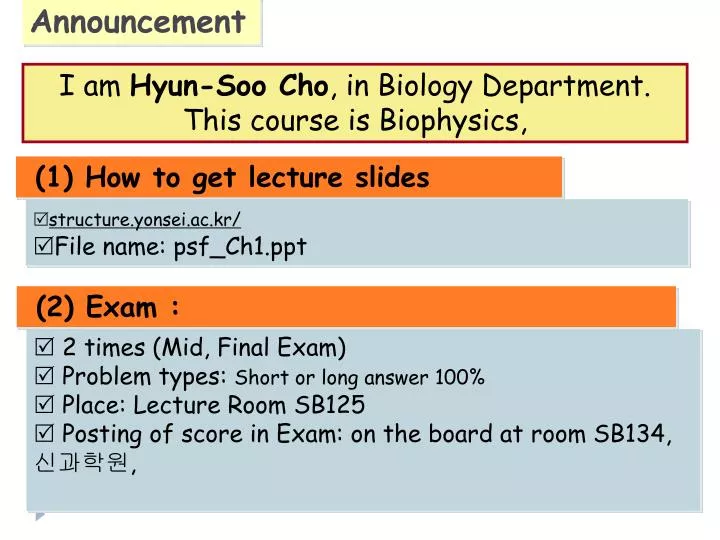
Announcement I am Hyun-Soo Cho, in Biology Department. This course is Biophysics, (1) How to get lecture slides • structure.yonsei.ac.kr/ • File name: psf_Ch1.ppt (2) Exam : • 2 times (Mid, Final Exam) • Problem types: Short or long answer 100% • Place: Lecture Room SB125 • Posting of score in Exam: on the board at room SB134, 신과학원,
Announcement (continued) (3) Assignment (homework): • Please read your textbook before or/and after each class (4) Grading: • Mid exam (35%) + Final exam (35%) + Presentation (15%) + attendance (5%) + Quiz (10%) (5) Participating in this Biophysics Course • Thanks everyone for your interest on this class • Have good manners : No cell phone (no message), No chatting • I hope you would keep your honor during this course
Announcement (continued) (6) Interviewing with me • You must see me during this course at least one time • My office hours: AM 10:00-12:00 on Friday • How: First, Contact me by E-mail or telephone • E-mail address: hscho8@yonsei.ac.kr,2123-5651
1-0. Overview : Protein Function and Architecture Figure 1-1. Four examples of biochemical functions performed by proteins
1-0. Overview : Protein Function and Architecture Figure 1-1. Four examples of biochemical functions performed by proteins
1-0. Overview : Protein Function and Architecture Figure 1-1. Four examples of biochemical functions performed by proteins
1-0. Overview : Protein Function and Architecture Sheet & strand Figure 1-1. Four examples of biochemical functions performed by proteins
1-0. Overview : Protein Function and Architecture Figure 1-2. Levels of protein structure illustrated by the catabolite activator protein
1-1. Amino Acids Figure 1-3. Amino-acid structure and the chemical characters of the amino-acid side chains
Structure and Stereoisomerism of a-Amino Acids COO- : carboxyl group Ca : a-carbon (chiral) NH3+ : amino group R : functional group (side chain) Absolute Configuration : S Left, Counter-Clockwise Absolute Configuration : R Right, Clockwise Only L-amino acids are constituents of proteins.
1-1. Amino Acids Figure 1-3. Amino-acid structure and the chemical characters of the amino-acid side chains
1-1. Amino Acids Figure 1-3. Amino-acid structure and the chemical characters of the amino-acid side chains
The amino-acid side chains have different tendencies to participate in interactions • Hydrophobic residues: van der Waals interactions – tendency to avoid contact with water and pack against each other hydrophobic effect - Ala & Leu are strong helix-favoring residues, Proline are not because its backbone nitrogen isn’t available for H-bond - Aromatic side chain of Phe participates in weakly polar interactions • Hydrophilic residues : Hydrogen bonds to one another, to peptide backbone, to polar organic molecules, and to water. - pKa shift: Asp & Glu (57 in hydrophobic interior or nearby (-) charge), Lys (106 in ?) - His: most versatile, most often found in enzyme active sites, pKa is 6, neutral, proton donator and acceptor
- Arg: completely protonated at neural, compared to Lys? - Cys: common in enzyme active site, most powerful nucleophile. Compared to Ser? • Amphipatic residues : both polar and nonpolar character - Lys: hydrophic charged region, long hydrophobic region (methylene) involved in van der Waals interactions with hydrophobic side chains - Tyr: pKa is 9, in some enzyme active site, hydroxyl group can be donor and acceptor of H-bond. Aromatic ring can form weakly polar interactions - Trp: similar to Tyr, indole amide hydrogen don’t ionize. - Met: least polar among amphipatic residues, thioether sulfur is excellent ligand for metal ions.
van der waals interaction - caused by transient dipoles, the momentary random fluctuation in the distribution of the electrons of any atoms
1-2. Genes and Proteins The genetic code is degenerate Figure 1-4. The genetic code
1-2. Genes and Proteins Splicing And ? Figure 1-5. The flow of genetic information in prokaryotes (left) and eukaryotes (right)
Alternative splicing can lead to truncated proteins, proteins with different stretches in the middle, and frameshifts. • Coding sequences can also be modified by RNA editing; some nucleotides can be changed and additional nucleotides inserted into the mRNA sequence before translation.
Genetic code organization • Single-base changes (single-nucleotide polymorphism) in the third position in a codon produce the same amino acid. Changes elsewhere in the codon produce a different amino acid, but with the same physical-chemical propherties. The second base specifies if the amino acid is polar or hydrophobic. Conservative substitutions.
1-2. Genes and Proteins Figure 1-6. Table of the frequency with which one amino acid is replaced by others in amino-acid sequences of the same protein from different organisms
1-3. The Peptide Bond Figure 1-7. Peptide bond formation and hydrolysis
1-3. The Peptide Bond • Resonance of peptide bond • Polarity, Dipole moment • partial double-bond character Figure 1-8. Schematic diagram of an extended polypeptide chain
1-3. The Peptide Bond Ramachandran plot Figure 1-9. Extended polypeptide chain showing the typical backbone bond lengths and angles
1-4. Bonds that Stabilize Folded Proteins Folded proteins are stabilized mainly by weak noncovalent interactions 1 kcal = 4.2 kJ Figure 1-10 Table of the typical chemical interactions that stabilize polypeptides
1-5. Importance and Determinants of Secondary Structure Peptide back bone C=O and N-H tend to hydrogen bond with one another, which result in the secondary structure. Especially in the interior of proteins. Figure 1-11. Ramachandran plot
: the angle of rotation about the bond between the nitrogen and the a-carbon y : the angle of rotation about the a-carbon and the carbonyl carbon Rotational Properties of Peptide Bonds Peptide bonds are rigid… But, the bonds containing the a-carbon between two peptide bonds can be rotated from -180o to +180o.
1-5. Importance and Determinants of Secondary Structure Prediction of secondary structure elements from a. a sequence is accurate to only about 70%. convenient way of fold classification N+3 beta turn, reverse turn, hairpin turn N Figure 1-12. Typical beta turn
1-6. Properties of the Alpha Helix N N+4 Figure 1-13. The alpha helix
1-6. Properties of the Alpha Helix • Lipid bilayer thickness? 30A. To span the cell membrane, how long helix? • at least 20 residues long helix • All helices in real protein structures are right-handed. Why? • because of steric hindrance caused by L-configuration • Helix dipole increase with increasing length of the helix • At the N-terminal ends of helices negative side chain Figure 1-14. Table of helical parameters
1-6. Properties of the Alpha Helix Alpha helices can be amphipathic, with one polar and one nonpolar face Figure 1-15. View along the axis of an idealized alpha-helical polypeptide
1-6. Properties of the Alpha Helix 1) Special examples of a-helix • Collagen : bone, tendon, ligament and blood vessel • Every third residue, glycine (GlyXY)n, X & Y proline • Proline lacks N-H groups, • hydroxylation! Figure 1-16. The structure of collagen
Collagen: the most abundant protein of mammals, main fibrous component of skin, bone, tendon, cartilage, and teeth. (피부미용)
2) Special examples of a-helix Coiled-coil protein • Structural support for Cells and Tissues • a-keratin: left-handed superhelix of two right-handed a helices. • from wool & hair, intermediate filaments in cytoskeleton, muscle protein (myosin & tropomyosin) • Heptad repeats; Every seventh residue in each helix, Leu holds two helix by van der Waals interactions • disulfide bond crosslinks: fewer – flexible, more – harder (horns, claws etc)
1-7. Properties of the Beta Sheet Figure 1-17. The structure of the beta sheet
1-7. Properties of the Beta Sheet No Plain b-sheet Only twisted b-sheet. Why? Stability & integrity of b-sheetdepends on # of b-strands Figure 1-18. Two proteins that form a complex through hydrogen bonding between beta strands (the Rap-Raf complex, PDB 1gua)
1-7. Properties of the Beta Sheet b strands usually have a pronouced right-handed twist, due to steric effects arising from the L-amino acid configuration. Figure 1-19. Beta barrel ; closed cylinder, retinol-binding protein
1-8. Prediction of Secondary Structure A-helices prediction Is easier than b-sheet Figure 1-20. Table of conformational preferences of the amino acids
1-8. Prediction of Secondary Structure Figure 1-21. An example of secondary structure prediction
1-9. Folding The structure of a protein is directly determined by its primary structure Figure 1-22. Folding intermediates
1-9. Folding Competition between self-interactions and interactions with water drives Protein folding Figure 1-23. Highly simplified schematic representation of the folding of a polypeptide chain in water
Computational prediction of folding is not yet reliable • Ab initio method - Equilibrium conformation is the global free-energy minimum - potential energy parameter is accurate (H-bond, van der Waals etc) - key intermediates? - oligomerization can not be addressed although very many globular proteins are oligomeric.
The polar amide and carbonyl group should hydrogen bond to one another because water can’t involve in H-bonds The hydrophobic environment of a membrane permits only all-helical and all-beta-barrel integral membrane
1-10. Tertiary Structure The condensation of multiple secondary structural elements leads to tertiary structure • Two proteins with similar secondary structure elements but different tertiary structures Figure 1-24. Comparison of the structures of triosephosphate isomerase and dihydrofolate reductase
1-10. Tertiary Structure - loops Found at the surface of protein and Exposed to the solvent Sites for protein recognition, ligand Binding and membrane interaction Often mutation sites without changing the core structure Often move as rigid bodies because their side chain pack together Figure 1-25. Variable loops
1-10. Tertiary Structure • Protein crystals contain more than 50% waters in their volumn • hydration shell • a few water inside the protein makes important interactions as • part of the tertiary structure Figure 1-26. Porcine pancreatic elastase showing the first hydration shell surrounding the protein
1-10. Tertiary Structure The atoms are packed as closely as in a solid. A few cavities and small channels provide some flexibility. Packing by ionic bonds, H-bonds, and van der Waal interactions. Packing types Figure 1-27. Cut-away view of the interior of a folded protein