Download
1 / 45
650 likes | 1.35k Views
osteoarthritis. Esmaeili ~ Esquivel ~ Fernandez ~ Ferrandiz ~ Flores ~ Francisco ~ Gansatao ~ Gatmaitan ~ Golpeo ~ Gutierrez. Approach to musculoskeletal complaint. Articular or Non-Articular?. < or > 6 Weeks?. Inflammatory or Not?. Which joints?. Articular vs non-articular.

E N D
osteoarthritis Esmaeili ~ Esquivel ~ Fernandez ~Ferrandiz ~ Flores ~ Francisco ~ Gansatao ~ Gatmaitan ~ Golpeo ~ Gutierrez
Approach to musculoskeletal complaint Articular or Non-Articular? < or > 6 Weeks? Inflammatory or Not? Which joints?
Articular vs non-articular Non-Articular Structures • Extra-articular ligaments • Tendons • Bursae • Muscle • Fascia • Bone • Nerve • Overlying skin Articular Structures • Synovium • Synovial Fluid • Articular cartilage • Intraarticular ligaments • Joint capsule • Juxtaarticular bone
Articular vs non-articular Features of Non-Articular • Point or focal tenderness • Painful of active ROM • Seldom demonstrate swelling, crepitation, instability or deformity Features of Articular • Deep or diffuse Pain • Pain or Limited ROM on active and passive movement • Swelling • Crepitation • Instability • “Locking” • Deformity
Approach to musculoskeletal complaint Articular or Non-Articular? < or > 6 Weeks? Inflammatory or Not? Which joints?
Inflammatory vs non-inflammatory • Cardinal signs of inflammation • Systemic symptoms • Laboratory evidence • Prolonged morning stiffness
Approach to musculoskeletal complaint Articular or Non-Articular? < or > 6 Weeks? Inflammatory or Not? Which joints?
OSTEOARTHRITIS Hip, DIP, PIP Articular Chronic Non-inflammatory
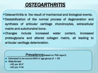
Osteoarthritis • Represents failure of the diarthrodial joint. • The most common joint disease in humans • Joint failure due to impaired joint protective mechanisms
Joint Failure • Joint failure occurs in the setting of loss of protective mechanisms • Joint protectors include: • Joint capsule and ligaments, synovial fluid • Muscle • Sensory afferents • Bone
Classification • Idiopathic • Localized • Hands • Feet • Knee • Hip • Spine • Other single sites, e.g., glenohumoral, acromioclavicular, tibiotalar, sacroiliac, temporomandibular • Generalized includes 3 or more of the area listed above
Classification • Secondary • Trauma • Congenital or developmental • Metabolic • Endocrine • Calcium deposition diseases • Neuropathic • Endemic • Miscellaneous • Frosbite • Caisson’s disease • hmoglobinopathies
I. Systemic Risk Factors • Age – most powerful risk factor. 2% prevalence among women <45years, 30% among 45-64 years and 68% among >65 years. • Cartilage are less responsive to stimulus for synthesis of matrix. Muscles and joints are less responsive to incoming loading movement. • Sensory nerve impulses are also slowed down with age thus halting the feedback mechanism of mechanoreceptor
Sex – more common in older women, possibly due to loss of estrogen during menopause. • Hip OA more common in male • Interphalangeal and thumb base OA more in women • Genetics – a woman with mother and sister affected with interphalangeal OA is 2-3x at risk • Race – Hip OA is less common in Chinese than Caucasians. OA more in Native Americans than in Caucasians.
II. Intrinsic Joint Vulnerabilities • Congenital hip diseases such as Legg-Perthes disease increase focal stress to hip joints increasing susceptibility to OA later in life. • Knee anomalies and malalignment such as Varus and Valgus deformity.
III. Loading Factors • Obesity – most potent risk factor for hip and knee OA. There is a linear relationship between risk of OA and increase in weight. 5kg weight loss is associated with 50% risk reduction. • Repetitive joint use – among miners, farmers, and runners.
Pathogenesis • The biomaterial properties of the articular cartilage and subchondral bone are normal, but excessive loading of the joint causes the tissues to fail. • The applied load is reasonable but the material properties of the cartilage or bone are inferior. • Decrease in polypeptide mediators which regulates biosynthesis of PGs responsible for compressive stiffness of tissue and withstand load. • Increase in IL-1 leading to suppression of PG synthesis and inhibiting matrix repair.
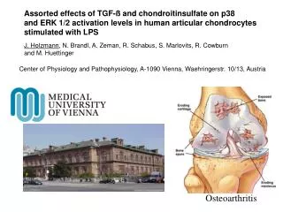
Pathogenesis • Hyaline cartilage loss. • Chrodrocytes attempts repair. Also stimulating inflammatory cytokine • Cartilage break down, bone exposure and development of subchondral cyst • Subchondral plate sclerosis, osteophyte growth and sinovitis. • Weakness of muscle bridging
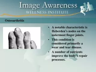
Clinical features • Joint stiffness / morning stiffness (<30 mins) • Joint pain (activity-related) • Episodic • Trigerred often by a day or two of overactive use of a diseased joint • Nocturnal pain • Mechanical symptoms: buckling, catching, or locking. • Limitation of joint movement • Deformity
Laboratory tests • No blood tests are routinely indicated • Examination of the synovial fluid is to rule out other causes of the pain and swelling.
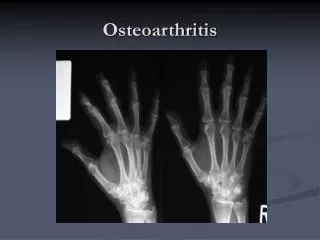
Imaging methods X-ray • Joint Space Narrowing • Development of Osteophytes • Subchondral Sclerosis • Subchondral Cyst Formation • Subluxation
Treatment goals • Pain reduction • Maintenance of mobility • Minimization of disability NICE Clinical Guideline 59
Pharmacologic treatment • Paracetamol first line drug for mild pain; max dose of 4g/day; close monitoring of upper GI adverse events • Tramadol control of moderate pain and improvement in knee function • Oral NSAIDs and COXIBs small to moderate effect in reducing exacerbations of knee OA; up to 2 weeks duration • Topical NSAIDs control of symptomatic or acute exacerbation of knee OA and improvement of function (PRA Practice Guidelines)
Pharmacologic treatment • Intraarticular Steroids effective and safe for moderate symptomatic exacerbations of knee OA; with effects up to 1-3 weeks (PRA Practice Guidelines) • Rubefacients / Capsaicin for reduction of joint pain and tenderness • Intraarticular Hyaluronic Acid for moderate pain and improvement in function; 3-5 weekly injections; longer duration of action than steroids (PRA Practice Guidelines)
Non-pharmacologic treatment Reduction of Joint Loading • Rest but not complete immobilization • Except in hand OA: DIP joint OA > custom-made splint to block flexion, improve overall hand function and reduce muscle spasm • Splinting Effective for trapeziometacarpal joint and pantrapezialOA
Non-pharmacologic treatment Patient Education • Provide additive benefit 20 to 30% as great as that of NSAID alone • Taking medications properly and communicating with health care providers • Decreases pain, disability and depression
Heat reduces pain and stiffness Hot shower or bath • Better analgesia with ice than heat • Wedged insoles / orthoses (polypropylene mesh insole = inexpensive and practical) - useful in OA of the medial tibialcompartment • Reduction of joint contact forces Unilateral OA cane should be held on contralateral side Bilateral Disease crutches or walker
Disuse of OA joint will lead to muscle atrophy • Periarticular muscles protects articular cartilage from stress hence strengthening exercises are important • Studies showed decreased pain, anxiety and depression with exercise • Weight loss for obese patients reduces joint loading 5% weight reduction significantly improves pain and function (PRA Practice Guidelines)
PATELLAR TAPING • Patellofemoral compartment severe pain Taping of patella reduces pain with isometric exercise to strengthen vastusmedialisobliquus component of quadriceps realignment of patella on a long term basis
ORTHOPEDIC SURGERY • Tidal irrigation of the knee • Arthroscopic debridement and lavage • Joint replacement for advanced OA • Joint arthroplasty may relieve pain and increase mobility • Osteotomy can eliminate concentration of peak dynamic loads and provides pain relief • Cartilage regeneration
references • Fauci, A.S et al (2008) Harrison’s Principles of Internal Medicine (17thed) NY: McGraw Hill Co, Inc. • National Institute for Health and Clinical Excellence (2008) “Osteoarthritis: The care and management of osteoarthritis is adults.” NICE Clinical Guideline 59. London: NHS. • Philippine Rheumatology Association (n.d.) “PRA Clinical Practice Guidelines for the Medical Management of Knee Osteoarthritis” Retrieved from http://philippinerheumatology.org/cgi-bin/news/news_details_print.asp?news_id=56 [07Aug 2011].