Download
1 / 35
510 likes | 1.17k Views
Osteoarthritis. Marcin Sibiński. Clinic of Orthopaedics and Paediatric Orthopaedics, Medical University of Lodz, Poland Prof. Marek Synder, M.D. Osteoarthritis (OA). 21 million people affected in USA 80-90% of individuals older than 65 years have evidence of primary osteoarthritis

E N D
Osteoarthritis Marcin Sibiński Clinic of Orthopaedics and Paediatric Orthopaedics, Medical University of Lodz, Poland Prof. Marek Synder, M.D.
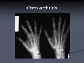
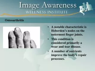
Osteoarthritis (OA) • 21 million people affectedin USA • 80-90% of individuals older than 65 years have evidence of primary osteoarthritis • OA of the knee and hips is the most common cause of arthritis-related disability • Commonly in the hands • Most common type of joint disease
Osteoarthritis (OA) • Middle-aged and older people • Age increases your risk for OA • Men under age 55 are more likely to have OA than women • After age 55, women are more commonly affected
Risk factors • Age • Obesity - for every kg. you gain, you add 3 kg. of pressure on your knees and 6 kg. the pressure on your hips • Injury or overuse (athletes) • Genetics • Muscle weakness • Other diseases (rheumatoid arthritis)
Pathophysiology • Hyaline cartilage (chondrocytes + extracellular matrix) • Protects the underlying subchondral bone • Distribution of large loads • Primarily begin in the articular cartilage • External forces accelerate the catabolic effects of the chondrocytes and disrupt the cartilaginous matrix
Pathological Features • Osteoarthritis represents a gradual processes of destruction & regeneration; • Early articular cartilage loses its glistening appearance; • Cartilage becomes thin and sometimes denuded;
Pathological Features • subchondral bone: • becomes thickened, sclerotic, & polished; • subchondral bone displays thickened trabeculae and microfractures; • cysts: • may be seen in subchondral bone; • cysts may arises from increases in intrasynovial pressure; • osteophytes: • spurlike bony outgrowths covered by hyaline cartilage, may develop at margins of joint & progressively enlarge; • small bits of cartilage-covered bone (mice) may actually break off into the joint;
Etiology - hip • Primary • associated with aging and is thought of as “wear and tear” • Secondary (predisposing condition) • Trauma (fractures) • Slipped capital femoral epiphysis • Developmental dislocation of the hip • Perthes disease Inflamatory arthritis (RA) • Osteonecrosis • Intra-articular steriods • Hyperparathyroidysa • Skeletal dysplasia • Hemochromatosis
Clinical Features - Hip • Pain on weitht-bearing felt in the groin, buttock, or medial thigh; • Pain during sleep; • Limping, ↓ROM • Trendelenburg gait will decrease mechanical stress on joint and thereby lessen pain; • In some cases, a patient with OA of the hip will experience acute hip pain which often correlates w/ rupture of subcondral cyst into the joint;
XR • Joint space narrowing • Osteophytes • Subchondral cyst formation • Subchondral sclerosis
Treatment • Non-operative treatment • Operative treatment: • Arthrodesis • Total hip replacement • valgus extension osteotomy
Goals of OA treatment • Controlling pain and other symptoms • Improving your ability to function in daily activities • Slow the disease’s progress
Non operative treatment • Keep fit • Non-steroidal anit-inflammatory drugs • Physical and occupational therapy
Non operative treatment • Injectable steroids • Viscosupplementation (hylauronic acid ) • Reduction of cartilage impact loading; • Cane, crutches • rubber heel wedges; • weight loss; • valgus unloading knee brace;
Operative treatment • Arthroscopy; • High tibial osteotomy; • Unicompartmental knee arthroplasty; • Total knee arthroplasty • Arthrodesis.