Download

1 / 53

E N D
Osteoarthritis Rowa’ Al Ramahi
DEFINITION • Osteoarthritis (OA) is a common, slowly progressive disorder affecting primarily the weight-bearing diarthrodial joints of the peripheral and axial skeleton. It is characterized by progressive deterioration and loss of articular cartilage, resulting in osteophyte formation, pain, limitation of motion, deformity, and progressive disability. Inflammation may or may not be present in the affected joints.
PATHOPHYSIOLOGY • Primary (idiopathic) OA, the most common type, has no known cause. Subclasses of primary OA are • localized OA (involving one or two sites) and • generalized OA (affecting three or more sites). • The term erosive OA indicates the presence of erosion and marked proliferation in the proximal and distal interphalangeal (PIP and DIP) hand joints
Secondary OA is associated with a known cause such as rheumatoid or another inflammatory arthritis, trauma, metabolic or endocrine disorders, and congenital factors. • OA usually begins with damage to articular cartilage through injury, excess joint loading from obesity or other reasons, or joint instability or injury that causes abnormal loading.
Damage to cartilage increases the metabolic activity of chondrocytes in an attempt to repair the damge, this leads to increased synthesis of matrix constituents with cartilage swelling. The normal balance between cartilage breakdown and resynthesis can be lost with a shift toward increasing destruction and cartilage loss. • After the hypertrophic phase, there is increased synthesis of matrix metalloproteinases (MMPs) 1, 3, 13, and 28, which causes collagen destruction to occur faster than synthesis, with a net loss of cartilage.
Chondrocytes contribute to collagen loss by secreting MMPs in response to inflammatory mediators present in OA (interleukin-1 and tumor necrosis factor-α). • Chondrocytes also undergo apoptosis, probably as a result of induction of nitric oxide synthase and production of toxic metabolites. This leaves fewer chondrocytes to synthesize matrix components. OA chondrocytes are also less responsive to the anabolic stimulus of transforming growth factor-β. The net result of these processes is a progressive cycle of cartilage destruction and chondrocyte loss.
Subchondral bone adjacent to articular cartilage also undergoes pathologic changes that allow progression of damage to articular cartilage. In OA, subchondral bone releases vasoactive peptides and MMPs. Neovascularization and subsequent increased permeability of the adjacent cartilage occur, which contribute further to cartilage loss. • Substantial loss of cartilage causes joint space narrowing and leads to painful, deformed joints.
The remaining cartilage softens and develops fibrillations, and there is splitting, further cartilage loss, and exposure of underlying bone. Cartilage is eventually eroded completely, leaving denuded subchondral bone that becomes dense, smooth, and glistening (eburnation). A more brittle, stiffer bone results, with decreased weight-bearing ability and development of sclerosis and microfractures. New bone formations (osteophytes) that arise from local and humoral factors appear at joint margins distant from cartilage destruction; evidence indicates that osteophytes help to stabilize OA joints.
Local inflammatory changes occur in the joint capsule and synovium. The synovium becomes infiltrated with T cells, and immune complexes appear. Crystals or cartilage shards in synovial fluid may contribute to inflammation. There are also increased levels of interleukin-1, prostaglandin E2, tumor necrosis factor- α, and nitric oxide in synovial fluid. Inflammatory changes result in effusions and synovial thickening. • The pain of OA arises from activation of nociceptive nerve endings within joints by mechanical and chemical irritants. OA pain may result from distension of the synovial capsule by increased joint fluid; microfracture; periosteal irritation; or damage to ligaments, synovium, or the meniscus.
CLINICAL PRESENTATION • The prevalence and severity of OA increase with age. Potential risk factors include obesity, repetitive use through work or leisure activities, joint trauma, and heredity. • The clinical presentation depends on duration and severity of disease and the number of joints affected. The predominant symptom is a localized deep, aching pain associated with the affected joint. Early in OA, pain accompanies joint activity and decreases with rest. With progression, pain occurs with minimal activity or at rest.
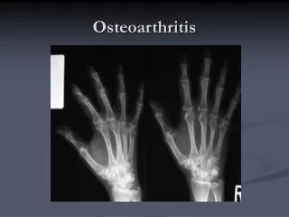
Joints most commonly affected are the DIP and PIP joints of the hand, the first carpometacarpal joint, knees, hips, cervical and lumbar spine, and the first metatarsophalangeal joint of the toe. • In addition to pain, limitation of motion, stiffness, crepitus, and deformities may occur. Patients with lower extremity involvement may report a sense of weakness or instability.
Upon arising, joint stiffness typically lasts less than 30 minutes and resolves with motion. Joint enlargement is related to bony proliferation or to thickening of the synovium and joint capsule. The presence of a warm, red, and tender joint may suggest an inflammatory synovitis.
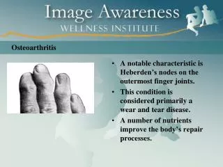
Joint deformity may be present in the later stages as a result of subluxation, collapse of subchondral bone, formation of bone cysts, or bony overgrowths. • Physical examination of the affected joints reveals tenderness, crepitus, and possible joint enlargement. Heberden and Bouchard nodes are bony enlargements (osteophytes) of the DIP and PIP joints, respectively.
DIAGNOSIS • The diagnosis of OA is dependent on patient history, clinical examination of the affected joint(s), radiologic findings, and laboratory testing. • Criteria for the classification of OA of the hips, knees, and hands were developed by the American College of Rheumatology (ACR). The criteria include the presence of pain, bony changes on examination, a normal erythrocyte sedimentation rate (ESR), and radiographs showing characteristic osteophytes or joint space narrowing.
For hip OA, a patient must have hip pain and two of the following: (1) an ESR less than 20 mm/hour, (2) radiographic femoral or acetabular osteophytes, or (3) radiographic joint space narrowing. • For knee OA, a patient must have knee pain and radiographic osteophytes in addition to one or more of the following: (1) age greater than 50 years, (2) morning stiffness of 30 minutes’ or less duration, or (3) crepitus on motion, (4) bony enlargement, (6) bony tenderness, or (7) palpable joint warmth.
No specific clinical laboratory abnormalities occur in primary OA. The ESR may be slightly elevated in patients with generalized or erosive inflammatory OA. The rheumatoid factor test is negative. Analysis of the synovial fluid reveals fluid with high viscosity. This fluid demonstrates a mild leukocytosis (less than 2,000 white blood cells/mm3) with predominantly mononuclear cells.
DESIRED OUTCOME • The major goals for the management of OA are to • (1) educate the patient, family members & caregivers • (2) relieve pain and stiffness; • (3) maintain or improve joint mobility; • (4) limit functional impairment; and • (5) maintain or improve quality of life.
TREATMENT • NONPHARMACOLOGIC THERAPY • The first step is to educate the patient about the extent of the disease, prognosis, and management approach. Dietary counseling and a structured weight-loss program are recommended for overweight OA patients. • Physical therapy—with heat or cold treatments and an exercise program— helps to maintain and restore joint range of motion and reduce pain and muscle spasms.
Exercise programs using isometric techniques are designed to strengthen muscles, improve joint function and motion, and decrease disability, pain, and the need for analgesic use. • Assistive and orthotic devices such as canes, walkers, braces, heel cups, and insoles can be used during exercise or daily activities. • Surgical procedures (e.g., osteotomy, partial or total arthroplasty, joint fusion) are indicated for patients with functional disability and/or severe pain unresponsive to conservative therapy.
PHARMACOLOGIC THERAPY • General Approach • Drug therapy in OA is targeted at relief of pain. Because OA often occurs in older individuals who have other medical conditions, a conservative approach to drug treatment is warranted. • An individualized approach to treatment is necessary. For mild or moderate pain, topical analgesics or acetaminophen can be used. If these measures fail or if there is inflammation, NSAIDs may be useful. Appropriate nondrug therapies should be continued when drug therapy is initiated.
Acetaminophen is recommended by the ACR as first-line drug therapy for pain management of OA. The dose is 325 to 650 mg every 4 to 6 hours on a scheduled basis (maximum dose 4 g/day; maximum 2 g/day if chronic alcohol intake or underlying liver disease). Comparable relief of mild to moderate OA pain has been demonstrated for acetaminophen (2.6 to 4 g/ day) compared with aspirin (650 mg four times daily), ibuprofen (1,200 or 2,400 mg daily), and naproxen (750 mg daily). However, some patients respond better to NSAIDs.
Acetaminophen is usually well tolerated, but potentially fatal hepatotoxicity with overdose is well documented. It should be used with caution in patients with liver disease and those who chronically abuse alcohol; when used in this setting, the duration should be limited & the dose should not exceed 2g/d. Chronic alcohol users (three or more drinks daily) should be warned about an increased risk of liver damage or GI bleeding with acetaminophen..
Other individuals do not appear to be at increased risk for GI bleeding. • Renal toxicity is also possible with long term use; use of non prescription combination products containing acetaminophen & NSAIDs is discouraged because of an increased risk of renal failure.
NSAIDs at prescription strength are often prescribed for OA patients after treatment with acetaminophen proves ineffective, or for patients with inflammatory OA. • Analgesic effects begin within 1 to 2 hours, whereas antiinflammatory benefits may require 2 to 3 weeks of continuous therapy.
Non selective NSAIDs & COX2 selective NSAIDs are superior to acetaminophen for improving symptoms & functional limitations in OA. • All NSAIDs have similar efficacy in reducing pain and inflammation in OA, although individual patient response differs among NSAIDs.
Selection of an NSAID depends on prescriber experience, medication cost, patient preference, toxicities, and adherence issues. An individual patient should be given a trial of one drug that is adequate in time (2 to 3 weeks) and dose. If the first NSAID fails, another agent in the same or another chemical class can be tried; this process may be repeated until an effective drug is found. Combining two NSAIDs increases adverse effects without providing additional benefit.
Cyclooxygenase-2 (COX-2) selective inhibitors (e.g., celecoxib) demonstrate analgesic benefits that are similar to traditional nonselective NSAIDs. Although COX-2 selective inhibition was designed to reduce NSAID induced gastropathy (e.g., ulcers, bleeding, perforation), concerns about adverse cardiovascular events (e.g., myocardial infarction, stroke) have led authorities to recommend their use only in selected patients who are at high risk for NSAID-related GI effects and low risk for cardiovascular toxicity. • •
GI complaints are the most common adverse effects of NSAIDs. Minor complaints such as nausea, dyspepsia, anorexia, abdominal pain, flatulence, and diarrhea occur in 10% to 60% of patients. • NSAIDs should be taken with food or milk, except for enteric-coated products (milk or antacids may destroy the enteric coating and cause increased GI symptoms in some patients).
All NSAIDs have the potential to cause gastric and duodenal ulcers and bleeding through direct (topical) or indirect (systemic) mechanisms. Risk factors for NSAID-associated ulcers and ulcer complications (perforation, gastric outlet obstruction, GI bleeding) include increased age, comorbid medical conditions (e.g., cardiovascular disease), concomitant corticosteroid or anticoagulant therapy, and history of peptic ulcer disease or upper GI bleeding.
For OA patients who need an NSAID but are at high risk for GI complications, the ACR recommendations include either a COX-2 selective inhibitor or a nonselective NSAID in combination with either a proton pump inhibitor or misoprostol. • NSAIDs may also cause kidney diseases, hepatitis, hypersensitivity reactions, rash, and CNS complaints of drowsiness, dizziness, headaches, depression, confusion, and tinnitus. All nonselective NSAIDs inhibit COX-1-dependent thromboxane production in platelets, thereby increasing bleeding risk.
NSAIDs should be avoided in late pregnancy because of the risk of premature closure of the ductus arteriosus. • The most potentially serious drug interactions include the concomitant use of NSAIDs with lithium, warfarin, oral hypoglycemics, high-dose methotrexate, antihypertensives, angiotensin-converting enzyme inhibitors, β- blockers, and diuretics.
Topical Therapies • Capsaicin, an extract of red peppers that causes release and ultimately depletion of substance P from nerve fibers, has been beneficial in providing pain relief in OA when applied topically over affected joints. It may be used alone or in combination with oral analgesics or NSAIDs.
To be effective, capsaicin must be used regularly, and it may take up to 2 weeks to work. It is well tolerated, but some patients experience burning &/or stinging at the site of application that usually subsides with repeated application. Patients should be warned not to get the cream in their eyes or mouth and to wash their hands after application. • Application of the cream, gel, or lotion is recommended four times daily, but tapering to twice-daily application may enhance long-term adherence with adequate pain relief.
Topical NSAIDs can provide effective OA treatment while avoiding the serious adverse effects of systemic therapy. They may be considered when first-line agents fail, are contraindicated, or are poorly tolerated. The mechanism of action is thought to be related to local inhibition of COX-2 enzymes. Common adverse effects include pruritus, burning, pain, and rash at the site of application. Patients using topical products should avoid oral NSAIDs to minimize the potential for additive side
Topical diclofenac 1.5% solution (Pennsaid) and diclofenac 1% gel (Voltaren Gel) are applied four times daily according to labeling instructions. • Topical rubefacients containing methyl salicylate, trolamine salicylate, and other salicylates may have modest, short-term efficacy for treating acute pain associated with OA. Local skin reactions occur rarely.
Glucosamine and Chondroitin • Glucosamine and chondroitin are dietary supplements that were shown to stimulate proteoglycan synthesis from articular cartilage in vitro. Although their excellent safety profile makes them appealing for patients at high risk of adverse drug events, results of a large, well-controlled, clinical trials sponsored by National Institutes of Health demonstrated no significant clinical response to glucosamine alone, chondroitin alone, or combination therapy when compared to placebo across all patients.
In subgroup analyses, patients with moderate to severe knee pain showed a response to combination glucosamine–chondroitin therapy superior to placebo, but this finding did not reach the predetermined threshold for pain reduction. • Because of their relative safety, a trial of glucosamine–chondroitin may be reasonable in patients with moderate to severe knee OA considering alternatives to traditional OA treatment. However, two recent evidence based guidelines recommend against their use.
Dosing should be at least glucosamine sulfate 1,500 mg/day and chondroitin sulfate 1,200 mg/day in divided doses. • Glucosamine adverse effects are mild and include GI gas, bloating, and cramps; it should not be used in patients with shellfish allergies. The most common adverse effect of chondroitin is nausea.
Corticosteroids • Systemic corticosteroid therapy is not recommended in OA, given the lack of proven benefit and the well-known adverse effects with long-term use. • Intraarticular corticosteroid injections can provide relief, particularly when a joint effusion is present. Average doses for injection of large joints in adults are methylprednisolone acetate 20 to 40 mg or triamcinolone hexacetonide 10 to 20 mg.
After aseptic aspiration of the effusion and corticosteroid injection, initial pain relief may occur within 24 to 72 hours, with peak relief occurring after 7-10 d and lasting for 4 to 8 weeks. • The patient should minimize joint activity and stress on the joint for several days after the injection. Therapy is generally limited to three or four injections per year because of the potential systemic effects of the drugs and because the need for more frequent injections indicates poor response to therapy.
Hyaluronate Injections • High-molecular-weight hyaluronic acid is a constituent of synovial fluids that provides lubrication with motion and shock absorbency during rapid movements. Because the concentration and molecular size of endogenous hyaluronic acid decrease in OA, exogenous administration has been studied in an attempt to reconstitute synovial fluid and reduce symptoms.
Hyaluronic acid injections temporarily and modestly increase synovial fluid viscosity and were reported to decrease pain, but many studies were short term and poorly controlled with high placebo response rates. • Six intraarticular hyaluronic acid preparations are available for treating Knee pain associated with OA:
sodium hyaluronate (Hyalgan 20 mg/2 mL) once weekly for five injections • sodium hyaluronate (Euflexxa 20 mg/2 mL) once weekly for three injections • sodium hyaluronate (Supartz 25 mg/2.5 mL) once weekly for five injections • Hylan polymers (Synvisc 16 mg/2 mL) once weekly for three injections • Hylan polymers (Synvisc one 48mg/6mL) sigle injection with efficacy for up to 26 weeks • Hyaluronan (Orthovisc 30 mg/2 mL) once weekly for three injections
Injections are well tolerated, but acute joint swelling effusion & stiffness as well as local skin reactions (e.g., rash, ecchymoses, or pruritus) have been reported. • These products may be beneficial for OA of the knee that is unresponsive to other therapy, but they are expensive because treatment includes both drug and administration costs.
Opioid Analgesics • Low-dose opioid analgesics (e.g., oxycodone) may be useful for patients who experience no relief with acetaminophen, NSAIDs, intraarticular injections, or topical therapy. • They are particularly useful in patients who cannot take NSAIDs because of renal failure or cardiovascular disease, or for patients in whom all other treatment options have failed and who are at high surgical risk, precluding joint arthroplasty.
Opioid Analgesics • Low-dose opioids should be used initially, allowing a sufficient duration between dose increases to permit an assessment for efficacy & safety. • Sustained release compounds usually offer better pain control throughout the day & are used when immediate release opioids do not provide a sufficient duration of pain control.
Tramadol • Tramadol with or without acetaminophen has modest analgesic effects in patients with OA. It may also be effective as add-on therapy in patients taking concomitant NSAIDs or COX-2 selective inhibitors. As with opioids, tramadol may be helpful for patients who cannot take NSAIDs or COX-2 selective inhibitors.
Tramadol should be initiated at a lower dose (100 mg/day in divided doses) and may be titrated as needed for pain control to a dose of 200 mg/ day. It is available in a combination tablet with acetaminophen and as a sustained-release tablet. • Opioid-like adverse effects such as nausea, vomiting, dizziness, constipation, headache, and somnolence are common.