Download
1 / 1
10 likes | 93 Views
PROTEIN THIN FILM MACHINES Stefania Federici 1,2 , Giulio Oliviero 1 , Kimberly Hamad-Schifferli 2,3 and Paolo Bergese 1 1 Chemistry for Technologies Laboratory and INSTM, University of Brescia, Via Branze, 38, 25123 Brescia , IT.

E N D
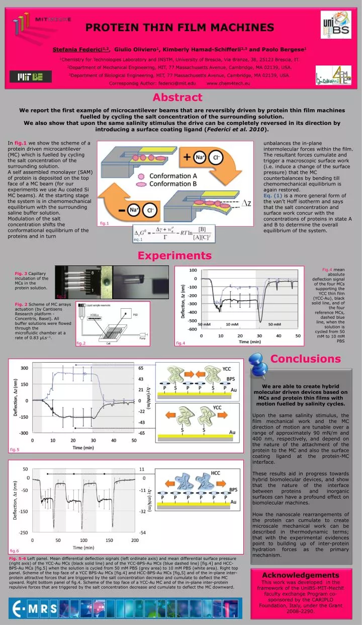
PROTEIN THIN FILM MACHINES Stefania Federici1,2, Giulio Oliviero1, Kimberly Hamad-Schifferli2,3 and Paolo Bergese1 1Chemistry for Technologies Laboratory and INSTM, University of Brescia, Via Branze, 38, 25123 Brescia, IT. 2Department of Mechanical Engineering, MIT, 77 Massachusetts Avenue, Cambridge, MA 02139, USA. 3Department of Biological Engineering, MIT, 77 Massachusetts Avenue, Cambridge, MA 02139, USA. CorrespondigAuthor: federici@mit.edu www.chem4tech.eu Abstract We report the first example of microcantilever beams that are reversibly driven by protein thin film machines fuelled by cycling the salt concentration of the surrounding solution. We also show that upon the same salinity stimulus the drive can be completely reversed in its direction by introducing a surface coating ligand (Federici et al. 2010). In fig.1 we show the scheme of a protein driven microcantilever (MC) which is fuelled by cycling the salt concentration of the surrounding solution. A self assembled monolayer (SAM) of protein is deposited on the top face of a MC beam (for our experiments we use Au coated Si MC beams). At the starting stage the system is in chemomechanical equilibrium with the surrounding saline buffer solution. Modulation of the salt concentration shifts the conformational equilibrium of the proteins and in turn unbalances the in-plane intermolecular forces within the film. The resultant forces cumulate and trigger a macroscopic surface work (i.e. induce a change of the surface pressure) that the MC counterbalances by bending till chemomechanicalequilibrium is again restored. Eq. (1) is a more general form of the van’t Hoff isotherm and says that the salt concentration and surface work concur with the concentrations of proteins in state A and B to determine the overall equilibriumof the system. fig.1 eq.1 Experiments Fig.4 mean absolute deflection signal of the four MCs supporting the YCC thin film (YCC-Au), black solid line, and of the four reference MCs, dashed blue line, when the solution is cycled from 50 mM to 10 mM PBS Fig. 3 Capillary incubation of the MCs in the protein solution. fig.3 Fig. 2 Scheme of MC arrays actuation (by CantisensResearch platform - Concentris, Basel). All buffer solutions were flowed through the microfluidic chamber at a rate of 0.83 μLs−1. fig.4 fig.2 Conclusions We are able to create hybrid molecular driven devices based on MCs and protein thin films with motion fuelled by salinity cycles. Upon the same salinity stimulus, the film mechanical work and the MC direction of motion are tunable over a range of approximately 90 mN/m and 400 nm, respectively, and depend on the nature of the attachment of the protein to the MC and also the surface coating ligand at the protein-MC interface. These results aid in progress towards hybrid biomolecular devices, and show that the nature of the interface between proteins and inorganic surfaces can have a profound effect on biomolecular machines. How the nanoscale rearrangements of the protein can cumulate to create microscale mechanical work can be described in thermodynamic terms; that with the experimental evidences point to building up of inter-protein hydration forces as the primary mechanism. fig.5 fig.6 Fig. 5-6 Left panel. Mean differential deflection signals (left ordinate axis) and mean differential surface pressure (right axis) of the YCC-Au MCs (black solid line) and of the YCC-BPS-Au MCs (blue dashed line) [fig.4] and HCC-BPS-Au MCs [fig.5] when the solution is cycled from 50 mM PBS (grey area) to 10 mM PBS (white area). Right top panel. Scheme of the top face of a YCC BPS-Au MCs [fig.4] and HCC-BPS-Au MCs [fig,5] and of the in-plane inter-protein attractive forces that are triggered by the salt concentration decrease and cumulate to deflect the MC upward. Right bottom panelof fig.4. Scheme of the top face of a YCC-Au MC and of the in-plane inter-protein repulsive forces that are triggered by the salt concentration decrease and cumulate to deflect the MC downward. Acknowledgements This work was developed in the framework of the UniBS-MIT-MechE faculty exchange Program co-sponsored by the CARIPLO Foundation, Italy, under the Grant 2008-2290.