Download
1 / 34
350 likes | 539 Views
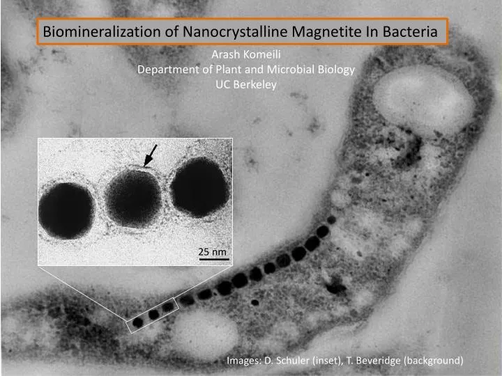
Biomineralization of Nanocrystalline Magnetite In Bacteria. Arash Komeili Department of Plant and Microbial Biology UC Berkeley. 25 nm. Images: D. Schuler (inset), T. Beveridge (background). Magnetotactic Bacteria. Morphologically and phylogenetically diverse

E N D
Biomineralization of Nanocrystalline Magnetite In Bacteria Arash Komeili Department of Plant and Microbial Biology UC Berkeley 25 nm Images: D. Schuler (inset), T. Beveridge (background)
Magnetotactic Bacteria • Morphologically and phylogenetically diverse • Orient in magnetic fields using magnetosomes • Use geomagnetic fields for direction-sensing
Magneto-aerotaxis • Frankel, Bazylinksi, Johnson and Taylor. Biophys Journal 1997 • Smith et al. Biophys Journal 2007
Why Study Magnetotactic Bacteria? -Geobiology “magnetofossils” as biomarkers -Applications Biomedicine, biotechnology, bioremediation -Biomineralization Genetic control of crystal properties D. Schuler
Magnetite Crystals are Formed Within Lipid Compartments 25 nm D. Schuler • Lipid Compartment • Unique set of proteins • Organized into chains with a cytoskeleton
Electron cryo-tomography of Magnetospirillum magneticum AMB-1 Dr. Grant Jensen Dr. Zhuo Li Division of Biology, California Institute of Technology Komeili, Li, Newman, Jensen. Science 2006
Model for Magnetosome Formation What are the genes and proteins that control these various functions?
Induction of Magnetite Synthesis +Fe -Fe
Genetic Screen Using a Magnetic Plate System 0 20 seconds 5 minutes
The Mutants WT mnm1 mnm2 mnm3 Grunberg et al. AEM (2001) mnm2 mnm3
Magnetosome Gene Island Contains the Majority of Magnetosome Genes Stats: 98 kb, 106 genes(or more!), 2% of the genome Fukuda et al. FEBS Letters, Feb. 2006
Genetic Dissection of the Magnetosome Island • Aims: • Identify genes involved in various steps of magnetosome formation • Define a minimum set of genes sufficient for magnetosome formation • Investigate the evolution and diversity of magnetosome formation Work by: Dr. Dorothee Murat
WT MaI 1 7 5 6 WT MaI 1 7 5 6 WT MaI 1 7 5 6 Anti-MamK Anti-MamA Anti-MamC mamABGene Cluster is a Central Regulator of Magnetosome Formation • Deletion of all 18 genes eliminates all traces of magnetosomes. • Next step: generate individual deletions of these 18 genes. • Each gene has a distinct role: • Membrane Formation • Chain Formation • Biomineralization
MamI, MamL, MamQ and MamB are essential for membrane biogenesis 0.2 µm DmamL strain H. Vali
Biomineralization Mutants mamV mutant mamS mutant What are the magnetic signatures of these mutants? ALS collaboration with Marco Liberati.
Model for magnetosome formation MamK, MamJ MamC, MamD, Mms6, MamA Komeili et al. PNAS 2004 Komeili et al. Science 2006 Scheffel et al. Nature 2006 Arakaki et al. JBC 2003
Model for magnetosome formation R2, Mam I, MamL, MamQ, MamB, MamE (?) ? MamK, MamJ MamC, MamD, Mms6, MamE, MamO MamMN, MamSTU, MamA, MamP R2, R3
Future Directions • Cell biological characterization of magnetosome formation • Identification of a minimum set of genes sufficient for magnetite formation • Evolution and diversity of magnetotactic bacteria
β-Proteobacteria γ-Proteobacteria α-Proteobacteria δ-Proteobacteria Burkholderia cepacia Shewanella alga Agrobacterium tumefaciens Desulfosarcina variabilis Magnetic vibrio MV-1 MMP Oceanospirillum pusillum Magnetospirillum AMB-1 Geobacter metallireducens Magnetospirillum MS-1 Desulfovibrio sp. BG6 MSM-3 and MSM-4 Desulfovibrio RS-1 Desulfovibrio sp. RS-1 Magnetospirillum MRS-1 Phaeospirillum moüsckianum Desulfovibrio desulfuricans Rhodospirillum rubrum Nitrospira moscoviensis Magnetococcus MC-1 Nitrospira CS92 Leptospirillum ferrooxidans CS103 Magnetobacterium bavaricum TB12 CS308 TB24 Cyanobacteria MP17 Itaipu I macpal19 macpal1 CS81 Planctomycetes macpal9 Thermotoga maritima Itaipu II Archaea Aquifex pyrophilus 10% tree from Amman et al. in Biomineralization 2004 One strain of magnetotactic bacteria outside the α-Proteobacteria has been cultured and sequenced Branches which contain magnetotactic bacteria (MTB) MTB which grow in culture and are sequenced
MS-1 MC-1 MSR-1 Taoka et al. 2006 Meldrum et al. 1993 AMB-1 RS-1 Arakaki et al. 2003 Grunberg et al. 2004 0.1 μm Desulfovibrio magneticus sp. RS-1 forms crystals with a different morphology Magnetite crystals from cultured α-Proteobacterial magnetotactic bacteria What cellular structures are involved in biomineralization of magnetite crystals in RS-1? Komeili et al. 2006
RS-1 forms round granules then magnetite crystals after addition of iron following iron starvation Number of electron-dense granules per cell Hours after iron addition At left are TEM images of whole RS-1 cells at different times after the addition of iron. At three hours, we begin to see the formation of round granules – which have not been seen previously in magnetotactic bacteria. By 22 hours, we begin to see magnetite crystals form, and by 50 hours the granules have disappeared and we see only magnetite crystals. At right, we have plotted the average number of granules and magnetite crystals per cell over time.
The granules are non-crystalline and contain iron and phosphate High-resolution TEM: magnetite crystals granules Elemental analysis (Energy-dispersive X-ray Spectoscopy) showed that the granules contain primarily iron and phosphorus.
Cryo electron microscopy shows membranes around iron-phosphate granules but not magnetite crystals Magnetite crystals Iron-phosphate granules 100 nm 100 nm Arrow highlights membrane.
Electron tomography of RS-1 thin section also shows no membranes around magnetite crystals • Conclusions: • Magnetite crystals in RS-1 do not seem to form within membrane-bounded compartments. This • suggests that RS-1 forms crystals using a distinct mechanism, perhaps a protein template. • RS-1 forms non-crystalline iron-phosphate granules within membrane-bounded compartments. • These compartments constitute a novel bacterial organelle.
Komeili Lab: Olga Draper Meg Byrne Dorothee Murat Anna Quinlan Shannon Greene Sepehr Keyhani Joyce Cueto Caltech: Dianne Newman Mel Simon Grant Jensen Zhuo Li Cody Nash McGill university: H. Vali • Funding: • David and Lucille Packard Foundation • Hellman Family Fund • NIH


![Nm]](https://cdn3.slideserve.com/6300766/slide1-dt.jpg)