Download
1 / 70
700 likes | 712 Views
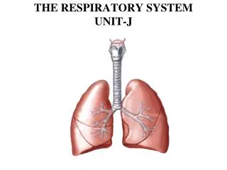
Unit 9 The Respiratory System. Function of the Respiratory System. Oversees gas exchanges (oxygen and carbon dioxide) between the blood and external environment Exchange of gases takes place within the lungs in the alveoli(only site of gas exchange, other structures passageways

E N D
Function of the Respiratory System • Oversees gas exchanges (oxygen and carbon dioxide) between the blood and external environment • Exchange of gases takes place within the lungs in the alveoli(only site of gas exchange, other structures passageways • Passageways to the lungs purify, warm, and humidify the incoming air • Shares responsibility with cardiovascular system
Function of the Respiratory System • Produces sound • Assists with the sense of smell • Assists in the regulation of blood pH
Organs of the Respiratory system • Nose • Pharynx • Larynx • Trachea • Bronchi • Lungs – alveoli
Anatomy of the Nasal Cavity • Olfactory receptors are located in the mucosa on the superior surface • The rest of the cavity is lined with respiratory mucosa • Moistens air • Traps incoming foreign particles
Anatomy of the Nasal Cavity • Lateral walls have projections called conchae • Increases surface area • Increases air turbulence within the nasal cavity • The nasal cavity is separated from the oral cavity by the palate • Anterior hard palate (bone) • Posterior soft palate (muscle)
Paranasal Sinuses • Cavities within bones surrounding the nasal cavity • Frontal bone • Sphenoid bone • Ethmoid bone • Maxillary bone
Paranasal Sinuses • Function of the sinuses • Lighten the skull • Act as resonance chambers for speech • Produce mucus that drains into the nasal cavity
Pharynx (Throat) • Muscular passage from nasal cavity to larynx • Three regions of the pharynx • Nasopharynx – superior region behind nasal cavity • Oropharynx – middle region behind mouth • Laryngopharynx – inferior region attached to larynx • The oropharynx and laryngopharynx are common passageways for air and food
Upper Respiratory Tract Figure 13.2
Structures of the Pharynx • Auditory tubes enter the nasopharynx • 5 inches long • Connects nose and mouth to the larynx and esophagus
Larynx (Voice Box) • Routes air and food into proper channels • Plays a role in speech • Made of eight rigid hyaline cartilages and a spoon-shaped flap of elastic cartilage (epiglottis)
Structures of the Larynx • Thyroid cartilage • Largest hyaline cartilage • Protrudes anteriorly (Adam’s apple) • Epiglottis • Superior opening of the larynx • Routes food to the larynx and air toward the trachea
Structures of the Larynx • Vocal cords (vocal folds) • Vibrate with expelled air to create sound (speech) • Glottis – opening between vocal cords
Trachea (Windpipe) • Connects larynx with bronchi • Lined with ciliated mucosa • Beat continuously in the opposite direction of incoming air • Expel mucus loaded with dust and other debris away from lungs • Walls are reinforced with C-shaped hyaline cartilage
Tracheal Obstruction • Kills over 4000 people each year • 5th major cause of accidental death in the US • Heimlich Maneuver is an emergency response to victims. http://www.youtube.com/watch?v=tEIiEAn7b-U
Primary Bronchi • Formed by division of the trachea into left and right primary bronchi • Enters the lung at the hilus (medial depression aka mediasternum- where the heart is located) • Right bronchus is wider, shorter, and straighter than left • Bronchi subdivide into smaller branches – Secondary bronchi and bronchioles
Lungs • Occupy most of the thoracic cavity • Apex is near the clavicle (superior portion) • Base rests on the diaphragm (inferior portion) • Each lung is divided into lobes by fissures • Left lung – two lobes • Right lung – three lobes
Coverings of the Lungs • Pulmonary (visceral) pleura covers the lung surface • Parietal pleura lines the walls of the thoracic cavity • Pleural fluid fills the area between layers of pleura to allow gliding
Respiratory Tree Divisions • Primary bronchi • Secondary bronchi • Tertiary bronchi • Bronchioli • Terminal bronchioli
Bronchioles • Smallest branches of the bronchi
Bronchioles • All but the smallest branches have reinforcing cartilage
Bronchioles • Terminal bronchioles end in alveoli
Respiratory Zone • Structures • Respiratory bronchioli • Alveolar duct • Alveoli • Site of gas exchange
Alveoli • Structure of alveoli • Alveolar duct • Alveolar sac • Alveolus Site of gas exchange
Respiratory Membrane (Air-Blood Barrier) • Thin squamous epithelial layer lining alveolar walls • Pulmonary capillaries cover external surfaces of alveoli
Gas Exchange • Gas crosses the respiratory membrane by diffusion • Oxygen enters the blood • Carbon dioxide enters the alveoli • Macrophages add protection • Surfactant coats gas-exposed alveolar surfaces
Events of Respiration • Pulmonary ventilation – moving air in and out of the lungs • External respiration – gas exchange between pulmonary blood and alveoli
Events of Respiration • Respiratory gas transport – transport of oxygen and carbon dioxide via the bloodstream • Internal respiration – gas exchange between blood and tissue cells in systemic capillaries
Mechanics of Breathing (Pulmonary Ventilation) • Completely mechanical process • Depends on volume changes in the thoracic cavity • Volume changes lead to pressure changes, which lead to the flow of gases to equalize pressure
Mechanics of Breathing (Pulmonary Ventilation) • Two phases • Inspiration – flow of air into lung • Expiration – air leaving lung
Inspiration • Diaphragm and intercostal muscles contract • The size of the thoracic cavity increases • External air is pulled into the lungs due to an increase in intrapulmonary volume
Exhalation • Largely a passive process which depends on natural lung elasticity • As muscles relax, air is pushed out of the lungs • Forced expiration can occur mostly by contracting internal intercostal muscles to depress the rib cage
Nonrespiratory Air Movements • Can be caused by reflexes or voluntary actions • Examples • Cough and sneeze – clears lungs of debris • Laughing • Crying • Yawn • Hiccup
Respiratory Volumes and Capacities • Normal breathing moves about 500 ml of air with each breath (tidal volume [TV]) • Many factors that affect respiratory capacity • A person’s size • Sex • Age • Physical condition • Residual volume of air – after exhalation, about 1200 ml of air remains in the lungs
Respiratory Volumes and Capacities • Inspiratory reserve volume (IRV) • Amount of air that can be taken in forcibly over the tidal volume • Usually between 2100 and 3200 ml • Expiratory reserve volume (ERV) • Amount of air that can be forcibly exhaled • Approximately 1200 ml
Respiratory Volumes and Capacities • Residual volume • Air remaining in lung after expiration • About 1200 ml
Respiratory Volumes and Capacities • Vital capacity • The total amount of exchangeable air • Vital capacity = TV + IRV + ERV • Dead space volume • Air that remains in conducting zone and never reaches alveoli • About 150 ml
Respiratory Volumes and Capacities • Functional volume • Air that actually reaches the respiratory zone • Usually about 350 ml • Respiratory capacities are measured with a spirometer
Respiratory Sounds • Sounds are monitored with a stethoscope • Bronchial sounds – produced by air rushing through trachea and bronchi • Vesicular breathing sounds – soft sounds of air filling alveoli
External Respiration • Oxygen movement into the blood • The alveoli always has more oxygen than the blood • Oxygen moves by diffusion towards the area of lower concentration • Pulmonary capillary blood gains oxygen
External Respiration • Carbon dioxide movement out of the blood • Blood returning from tissues has higher concentrations of carbon dioxide than air in the alveoli • Pulmonary capillary blood gives up carbon dioxide • Blood leaving the lungs is oxygen-rich and carbon dioxide-poor
Gas Transport in the Blood • Oxygen transport in the blood • Inside red blood cells attached to hemoglobin (oxyhemoglobin [HbO2]) • A small amount is carried dissolved in the plasma
Gas Transport in the Blood • Carbon dioxide transport in the blood • Most is transported in the plasma as bicarbonate ion (HCO3–) • A small amount is carried inside red blood cells on hemoglobin, but at different binding sites than those of oxygen
Internal Respiration • Exchange of gases between blood and body cells • An opposite reaction to what occurs in the lungs • Carbon dioxide diffuses out of tissue to blood • Oxygen diffuses from blood into tissue