Download
1 / 29
290 likes | 403 Views
Comparative enrichment of Phosphopeptides from ergosterol-treated A.thaliana leaves. Robyn Klemptner University of Johannesburg MSc supervisors: Dr. L.A. Piater Prof. I.A. Dubery Prof. R. Meijboom. Background. BIGGEST CHALLENGE : 9 BILLION people by 2050!!!

E N D
Comparative enrichment of Phosphopeptides from ergosterol-treated A.thaliana leaves Robyn Klemptner University of Johannesburg MSc supervisors: Dr. L.A. Piater Prof. I.A. Dubery Prof. R. Meijboom
Background • BIGGEST CHALLENGE: 9 BILLION people by 2050!!! • Food security – global importance. • Plant exposed to multiple pathogens. • Price hikes – plant diseases. • Preformed defenses. • Innate immunity = overcome pathogens. • PAMP-Triggered Immunity (PTI) + Effector-triggered immunity (ETI). (Lochman & Mikes, 2006 ; Godfray, 2009)
Innate immune responses • MAMPs/PAMPs • Preformed defenses compromised. • Bind PRR at cell membrane. • Signal transduction. • WRKYs. • MAMP/PAMP-triggered immunity (M/PTI). • Effectors • Against specific host. • Suppress M/PTI. • Effector-triggered immunity (ETI). • Recognized by intracellular receptors. • ROS, HR, SAR. Figure 1: A figure that clearly indicates the two mechanisms of pathogen detection and induction of corresponding immune responses. (Klemptner et al., 2014)
Ergosterol – an “orphan” MAMP • Ergosterol = Fungal sterol, fungal cell membrane component. • Implicated in major crop losses world wide. • Receptor/signal transduction pathway not yet elucidated. • Trigger immune response in sugar beet, grape, tomato and tobacco plants. • Reactive oxygen species, ion fluxes, PR proteins, LTPs. A B C D E F Figure 2: 3D models of various sterol compounds that have been used to study receptor interactions in plant-pathogen interactions. A: Ergosterol; B: Brassicasterol; C: Sitosterol; D: Stigmasterol; E: Campesterol; F: Cholesterol. (Avrovaet al., 2004; Wang , 2004; Rossard et al., 2010; Weeteet al., 2010; Klemptner et al., 2014)
What we know…. • Calcium-dependent protein kinases – Ca2+ influx. • Phospholipase Kinase C. • MAPKs. • WRKY transcription factors. • Phenylpropanoid pathway – metabolites. • H2O2 generation. • Ergosterol perception is specific.
Phosphorylation = Post-translational modification = structural change = functional change Serine, Threonine and Tyrosine residues of proteins = kinases = signal transduction activation. KinasesvsPhosphatases= regulation. (Schulze, 2010)
Phosphoproteins & signal transduction Figure 3: An overview of signal transduction pathways in defense responses in plants. (Yang et al, 1997; Thurston et al., 2005)
Enriching phosphoproteins • Important players in signal transduction BUT occur in low abundance! < only transiently phosphorylated! • Provide a greater knowledge of defense-related signal transduction networks. • Methods of enrichment include: • Affinity chromatography • Antibody-based affinity capture • Chemical derivatization • Metal ion-based affinity capture • Thus, more sensitive and reliable method required = DENDRIMERS! • Novel proteome investigation in plants since dendrimer-based enrichment techniques have yet to be applied to plant studies. (Meimounet al., 2007; Iliuk et al., 2010)
Dendrimers Figure 4: Dendrimer nanopolymers of varying generations. (Holisteret al., 2003)
Dendrimer isolation mechanism Add dendrimer to tryptic digest Filter through spin-column to isolate dendrimer + bound peptides Phosphorylated groups bind to surface amino groups Cleave peptides by acid hydrolysis Figure 5: The fundamental dendrimer-based phosphopeptide isolation mechanism. (Peters, 2005)
PolyMAC and PAMAM • Dendrimers with modified terminal groups on the surface. • Specific affinity for phosphorylated amino acid residues. A B Figure 6A & B: The PolyMAC dendrimer and its 2 types of side-chain moieties; the traditional PAMAM dendrimer with amine surface groups. (Iliuk et al., 2010; Mandeville & Tajmir-Raihi, 2010)
Objectives • Elicitation of A.thaliana with ergosterol and total protein expression profiles. • Enrich plant phosphopeptides using dendrimer technologies. • Compare efficiencies of PAMAM vs. PolyMAC dendrimer enrichment techniques. • Successful identification of differentially expressed phosphorylated proteins by Mass spectrometry. • Possibly elucidate ergosterol-induced signal transduction pathway of A. thaliana .
Methodology • PAMP treatment of A.thaliana plants • Untreated control • 250 nM ergosterol • EtOH control • 0, 6, 12, 24, 48, 72 hr and 7 days • SDS sample buffer • SDS-PAGE gels (1D) • Western blotting • Total protein extraction • Liquid N2 • TCA/acetone/phenol • Ammonium acetate/meOH precipitation • Buffers for downstream protocols • Urea sample buffer • PolyMAC and PAMAM enrichment • IEF sample buffer • Isoelectric focusing (2D) • Protein concentration quantification • Amido black assay • BSA standards (0.625, 1.25, 2.5, 5 and 10 ug/uL) • Samples and standards – nitrocellulose membrane • Absorbance at 600 nm (Granado, 1995; Lochman and Mikes, 2004; Wang et al., 2006)
Methodology • SDS-PAGE (1D) • 10 ug total/lane • 10% gel • Fairbanks/silver staining • Western Blotting • 1° Ab • = Anti-active MAPK • = Anti-phosphoTyr • IEF (2D-PAGE) • pH 3-10 and pH 4-7 • Fairbanks/silver staining • Dendrimer enrichment • Trypsin digest • C-18 peptide clean up • Enrichments • = PAMAM • =PolyMAC • Mass spectrometry analysis • MALDI-TOF • =DHB/CHCA • LC-MS/MS • Peptide sequences • Protein ID = MASCOT
SDS-PAGE: total protein kDA 260 140 100 70 50 40 35 25 15 10 EtOH EtOH EtOH Erg EtOH EtOH Erg Erg EtOH EtOH Erg Erg Erg Erg M UT M ~27 kDa 0hr 6hr 12hr 24hr 48 hr 72hr 7 days Figure 8: SDS-PAGE separation of all protein samples. Despite there being a large number of bands that are common to all the samples, there is a protein that shows differential expression and has an approximate size of 27 kDa.
Table 1: Protein identities following Mass Spectrometry of gel slices
A B pH 4 - 7 pH 4 - 7 C D pH 4 - 7 pH 4 - 7 Figure 9A, B, C & D: 2D-PAGE gels (11.25%) of ergosterol-treated samples following IEF,on a pH 4-7 IPG strip. Figure A shows spots resulting from the untreated control and those in figure B show those resulting from a 0 hour ergosterol treatment. Figures C and D show spots resulting from a 6 hr and 12 hr ergosterol treatment respectively.
Western Blotting – Anti phosphotyrosine ~40 kDa ~27 kDa UT 0hr 6hr 12hr 24hr 48 hr 72hr 7 days Figure 10: Autoradiography films showing Tyrosine-phosphorylated proteins following Western blotting. The dotted yellow boxes indicate a ~27 kDa protein that exhibits a strong binding signal to the anti-active phosphotyrosine antibody.
Western blotting – Anti active MAPK ~ 40 - 45 kDa ~ 15 - 25 kDa UT 0hr 6hr 12hr 24hr Figure 11: Autoradiography film showing the presence of MAPKs at 42 – 45 kDa.
MALDI-TOF mass spectrometry • Preliminary analysis of phosphopeptide enrichment. • DHB and CHCA matrices. • α-casein/BSA standard + samples + calibration peptides. • BrukerDaltonicsAutoFlex at the CSIR, Biosciences. • Nitrogen laser/ positive ion mode.
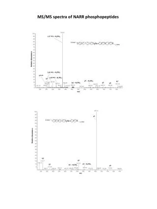
MALDI-TOF Figure 12: MALDI-TOF spectra of phosphopeptide standard (α-casein/BSA) and PolyMAC enriched sample.
Conclusions • Preliminary MALDI analysis indicates successful phosphopeptide enrichment. • Anti-PhosphoTyr = specific phosphoproteins. • ~27 kDa protein across samples = phosphorylated protein. Confirm identity. • Ergosterol-specific proteins = germin-like protein. • Defense and stress-related proteins are evident = aquaporins, LRR, calcium binding, Ras-related protein. (Klemptner et al., 2014)
Further studies and research outcomes • Final LC-MS/MS analysis = CSIR (Pretoria)/CPGR (Cape Town). • Identify total differentially expressed proteins. • Compare to western blots, SDS-PAGE and 2D. • Compare enrichment of in-gel digested proteins to proteins in solution – efficiency of dendrimer-based enrichments. • Compare genomic, proteomic and metabolomic data.
Acknowledgements • Dr. L. Piater, Prof. Dubery, Prof. R. Meijboom. • Prof. A.W. Tao – Tymora Analytical/ Purdue University – Indiana, USA. • National Research Foundation. • Dr. Stoyan Stoychev – CSIR Biosciences, Pretoria. • Dr. Salome Snyman– Stellenbosch University.
References Avrova, A.O., Taleb, N., Rokka, V-M., Heilbronn, J., Campbell, E., Hein, I., Gilroy, E.M., Cardle, L., Bradshaw, J.E., Stewart, H.E., Fakim, Y.J., Loake, G. and Birch, P.R.J. (2004) Potato oxysterol binding protein and cathepsin B are rapidly up-regulated in independent defense pathways that distinguish R-gene-mediated and field resistance to Phytophthora infestans. Molecular Plant Pathology, 5: 45-56. Boller T. and He Y.S, (2009) Innate Immunity in Plants: An Arms Race Between Pattern Recognition Receptors in Plants and Effectors in Microbial Pathogens. Journal of Science, 324: 742-744. Dodds, P. N., & Rathjen, J. P. (2010). Plant immunity: towards an integrated view of plant-pathogen interactions. Nature reviews. Genetics, 11 (8), 539-48. Fairbanks G, Steck TL, W. D. (1971). Electrophoretic analysis of the major polypeptides of the human erythrocyte membrane. Biochemistry, 10 (13), 2606-17. Godfray, H. C. J., Beddington, J. R., Crute, I. R., Haddad, L., Lawrence, D., Muir, J. F., Pretty, J., et al. (2010). Food security: the challenge of feeding 9 billion people. Science , 327 (5967), 812-8. Goldring, J. P., & Ravaioli, L. (1996). Solubilization of protein-dye complexes on nitrocellulose to quantify proteins spectrophotometrically. Analytical biochemistry, 242 (2), 197-201. Holister, P., Vas, C.R., Harper, T., (2003) Dendrimers. Clientifica, New York, pg 2-15. Iliuk, A. B., Martin, V. A, Alicie, B. M., Geahlen, R. L., & Tao, W. A. (2010). In-depth analyses of kinase-dependent tyrosine phosphoproteomes based on metal ion-functionalized soluble nanopolymers. Molecular & Cellular Proteomics , 9 (10): 2162-72. Klajnert, B., & Bryszewska, M. (2001). Dendrimers: properties and applications. Acta biochimica Polonica, 48 (1), 199-208. Klemptner, R.L., Sherwood, J. S., Tugizimana, F., Piater, L. A., & Dubery, I. A. (2014). Ergosterol, an orphan fungal microbe-associated molecular pattern (MAMP). Molecular Plant Pathology
Lochman J. and Mikes V., (2006) Ergosterol treatment leads to the expression of a specific set of defence-related genes in tobacco. Journal of Plant Molecular Biology, 62:43–51. Mandeville, J. S., & Tajmir-Riahi, H. A. (2010). Complexes of dendrimers with bovine serum albumin. Biomacromolecules, 11 (2): 465-72. Meimoun, P., Ambard-Bretteville, F., Colas-des Francs-Small, C., Valot, B., & Vidal, J. (2007). Analysis of plant phosphoproteins. Analytical biochemistry, 371(2): 238-46. Peters, E. C. (2005). A polymeric solution for enriching the phosphoproteome Insect transgenesis by site-specific. Nature Methods, 2(8): 579-580. Rossard S., Roblin G. and Atanassova R., (2010) Ergosterol triggers characteristic elicitation steps in Beta vulgarisleaf tissues. Journal of Experimental Botany, 61: 1807–1816. Schulze, W. X. (2010). Proteomics approaches to understand protein phosphorylation in pathway modulation. Current opinion in plant biology, 13(3): 280-87. Tao, W. A., Wollscheid, B., Brien, R. O., Eng, J. K., Li, X.-jun, Bodenmiller, B., Watts, J. D., et al. (2005). Quantitative phosphoproteome analysis using a dendrimer conjugation chemistry and tandem mass spectrometry. Nature Methods, 2(8): 591-598. Thurston G., Regan S., Rampitsch C., Xing T., (2005) Proteomic and phosphoproteomic approaches to understand plant–pathogen interactions. Journal of Physiological and Molecular Plant Pathology, 66: 3–11. Wang, W., Vignani, R., Scali, M., & Cresti, M. (2006). A universal and rapid protocol for protein extraction from recalcitrant plant tissues for proteomic analysis. Electrophoresis, 27(13): 2782-6. Weete J.D., Abril M., Blackwel M. (2010) Phylogenetic Distribution of Fungal Sterols. PLoS One. 5: 1-6. Yang Y., Shah J., and Klessig D.F. (1997) Signal perception and transduction in plant defence response. Journal of Genes and development,12: 1621-1628.