Download
1 / 45
450 likes | 927 Views
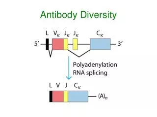
BASIC ANTIBODY IDENTIFICATION. Jean Purcelli, MT (ASCP)SBB Blood Centers of the Pacific May 2010 Version 2 May 2012. There are two types of unexpected red blood cell antibodies. Alloantibodies which may occur as a result from: Transfusion Pregnancy Transplantation

E N D
BASIC ANTIBODY IDENTIFICATION Jean Purcelli, MT (ASCP)SBB Blood Centers of the Pacific May 2010 Version 2 May 2012
There are two types of unexpected red blood cell antibodies. Alloantibodies which may occur as a result from: Transfusion Pregnancy Transplantation Injections of immunogenic material I.V. or I.M. material such as IVIg or RhIg (passively acquired) Autoantibodies which occur with certain diseases or medical treatments.
Alloantibodies can be detected in about 0.3%-5% of the general population. The incidence increases if the transfused or previous pregnancy populations are studied. The incidence is affected by: Antigenicity Prevalence of the antigen in the population Status of the immune system
Anti-A and Anti-B are examples of naturally occurring antibodies and they are expected. Lewis (Anti-Lea, Leb), -P1, -M, -N are unexpected antibodies, but can occur naturally. Naturally occurring antibodies have resulted from environmental, bacterial, or viral antigens that are similar to blood group antigens.
An Alloantibody is considered Clinically Significant: • If it has caused hemolytic transfusion reactions. • If it has caused marked decrease in the survival of transfused red cells. • If it has caused hemolytic disease of the fetus or newborn (HDFN).
We concentrate on identifying 13 clinically significant alloantibodies that are commonly found: Rh system antibodies: D C E c e f Kell system antibodies: K Duffy system antibodies: FyaFyb Kidd system antibodies: JkaJkb MNSs system antibodies: S s sometimes M (-M may be significant if it reacts at 37 C or by IAT)
Clinically Insignificant Several clinically insignificant alloantibodies and autoantibodies are commonly found. Alloanti-M (reacting only below 37ºC), -N, -P1, -Lea, -Leb, generally react below 37ºC and usually are not clinically significant. It is usual to transfuse with pre-warmed IAT, crossmatch compatible red cells. The IS crossmatch is dropped. Autoanti-I and –IH are commonly found when testing is conducted at room temperature or colder.
Red Cell Panels and Master Sheets A red cell panel is merely an expanded antibody detection set with 8-10 cells designed to identify and rule out the commonly found antibodies. Make copies of both sides of the Master Lists when they arrive, a new set will arrive every 2-4 weeks. The panel sheets are used as work sheets. The lot number of the panel should match the master sheet. Use in-dated cells whenever possible.
Panel Master Sheets The panel is designed to have some homozygous expressions of antigens, except P1. In addition K, Kpa, Jsa, and Lua antigens rarely exist as homozygous expressions on a panel. Next to the Vial Number is a column for Special Types. There are columns for four High Frequency antigens: k, Kpb, Jsb, Lub. Cells negative for these antigens rarely exist in panels. There is a note in the lower left listing several more high frequency antigens for which each cell was typed: I, Ge, Yta, Tja, Vel, Coa, Dib.
Panel Master Sheets There are columns for five Low Frequency antigens: V, Cw, Kpa, Jsa, Lua. In addition, there is a note in the lower left corner listing several more antigensof low frequency that each cell has been typed and found negative for Mg, Vw, Dia, Wra. On the reverse side is another list of Extended Typings and notations.
PRE-TESTING PHASE Obtain Medical and Transfusion History • Age: may affect ABO typing. • Sex • Ethic origin: high prevalence antigens are lacking in certain populations. • Diagnosis: some diseases are associated with some antibodies; warm and cold AIHA. • Transfusions and Pregnancies: how many transfusions, when, number of pregnancies
PRE-TESTING PHASE • Medications • Intravenous solutions • Check file for antibody history, difficulty in typing or transfusion reaction or HDFN
PLAN FOR TESTING PHASE Each institution should develop a plan for antibody identification. A plan establishes consistency and a general guide to antibody identification. An example plan: • Use a specifically designed worksheet, panel sheets, and selected cell panel sheets. • Obtain patient history. • Check files for previous work-up.
PLAN FOR TESTING PHASELab Testing • ABO-Rh • DAT • Initial Panel, use basic enhancement media such as LISS. • A cold antibody screening.
PLAN FOR TESTING PHASE • Establish identification and rule out other possible antibodies in PEG/IAT. • Perform partial or extended phenotype if there were no recent transfusions. • Other tests as necessary. Based on other findings. Steps 4-7 may be performed simultaneously.
TEST SYSTEM Most test systems include enhancement media and antiglobulin reagent. Testing with panel cells is conducted at 37ºC, examined for agglutination or hemolysis and converted to the antiglobulin test. A separate cold screen with Screening Cells and an auto control (in saline) offers valuable information.
THE ANALYTICAL PHASE Exclude specificities with negative reactions using a cross-out method. A single antibody usually produces a clear pattern. • Both positive and negative reactions are important. • Results should correspond with reactions found with the antibody screening cells. • Rule out other possible antibodies. • A selected cell panel may be necessary. • When the antibody(s) have been identified, the next step is to confirm the identity by typing the patient’s red cells for the antigens.
If the patient’s plasma is reactive with 2 cells positive for the antigen andnon-reactive with 2 cells negative for the antigen the minimum requirement for identification has been met. Additional reactive antigen +cells and non-reactive antigen = cells increase the probability of correct identification of the antibody.
To exclude other possible antibodies, at least two cells with a homozgous expression of the antigen should be non-reactive, whenever possible. The exclusion method by crossing out is a tentative and imperfect method to use until criteria have been met with a selected cell panel using the most sensitive enhancement media.
Rules for exclusion: Exceptions • Not all antigens exist as a homozygous expression. P1,Cw, V, VS. • Many low frequency antigens are not readily available as homozygous expressions: K, Jsa, Kpa, Lua, etc.
More exceptions to exclusion with homozygous cells: • Anti-C when anti-e is present. Conclusion: “cannot exclude possible anti-C” • Anti-E when anti-c is present. Conclusion: “cannot exclude possible anti-E”
More exceptions to exclusion with homozygous expression • Anti-C when anti-D is present. OK to exclude -C with r’r cells. • Anti-E when anti-D is present. OK to exclude -E with r”r cells.
A possible antibody can also be ruled out by typing for the antigen. If a person’s cells type positive for the antigen, that person will not make the corresponding antibody. Exceptions: There are many instances of D+ people producing anti-D and many auto-antibodies show specificities in the Rh system, particularly -e.
Monoclonal anti-Rh antisera show discrepant results when the antibody is made from different clones or sources. • A C+ person may make antibody appearing to have anti-C specificity. Is it an autoantibody or is the patient C negative? • It is advantageous to have more than 1 source of antisera.
In general a person who types negative for an antigen is capable of producing the antibody. Lewis system antibodies are an exception: Only Le(a-b-) persons can make Lewis antibodies of any specificity.
If no discernable pattern is observed consider: Multiple antibodies: D+C, or D+E, or E+K An antibody that demonstrates dosage: anti-M,-N,-S,-s,-Jka,-Jkb, -C, -E,-c,-e Some antigens have variable expression in different individuals: P1, I, “HTLA” Unwanted reactions from cold or warm autoantibodies. Refer to the DAT or autocontrol and the cold antibody screen for clues.
Multiple Antibodies There are several different approaches to resolving multiple antibodies : • Phenotyping • Selected Cell panel • Neutralization
Multiple antibodies • Enzymes • Adsorption We will concentrate on the most efficient way: a combination of phenotyping and using selected cell panels.
Multiple Antibodies • Extended phenotyping is expensive and should be used sparingly. • From a phenotype, you learn which antibodies are possible and which can be ruled out. • Then a selected cell panel is created and tested.
Selected Cell Panels • Use a selected panel worksheet. • Selected cells are chosen from other panel or screening cells to confirm or eliminate possible antibodies. • Chosen cells should be positive (+) for the suspected antigen and negative (0) for others.
Selected Cell Panels • Choose homozygous expressions whenever possible. • Record the (+) and (o) antigens and include the panel lot number, the donor number and the vial number for each cell chosen. • Test with PEG or LISS. Analyze results.
Proteolytic Enzymes Two stage techniques using either ficin or papain to pre-treat red cells are the most useful. Enzymes are used to enhance or destroy certain blood group antigens. Enzyme technique is useful in sorting out multiple antibodies, enhancing weak antibodies, providing clues to the identity of certain antibodies to high prevalence antigens, and can be used in certain adsorption procedures.
Proteolytic Enzymes Pre-treating panel cells takes about 10 minutes, then the serum is added and placed at 37º for 30 minutes. With papain, “pan-agglutination” is often seen at the 37º stage and is disregarded if it is seen in all cells, including the auto control. Unwanted reactions due to cold allo or autoantibodies and warm autoantibdies may be enhanced.
Antigens destroyed: M, N, S, Fya, Fyb. Small s is variable and should be QC’d with anti-s. Antigens enhanced: Rh: D, E, c, e, f, and all Rh antigens. Jka, Jkb. Lewis, I. Antigens unchanged: K, k and all Kell antigens.
DAT POSITIVE • The red cells were coated in vivo. • In a transfusion reaction, the transfused cells have become coated with immunoglobulins and the reactions can be quite weak. • In Warm Auto Immune Hemolytic Anemia, the red cells are coated with IgG only or with both IgG and C3, and the reactivity is often very strong. • In Cold Agglutinin Disease, the red cells are coated with C3. • In HDFN, the mother’s antibody has crossed the placenta and coated the fetus’ red cells.
IgG coated cells (positive DAT) cannot be typed with antisera requiring the antiglobulin test. • Monoclonal, direct agglutinating reagents reagents can be used reliably. • To test for Fya, Fyb, Jka, Jkb, S, s; the cells must be treated first with glycine-HCI-EDTA (EgA) to remove the bound IgG. • Glycine-HCI-EDTA destroys all Kell antigens. To type for K, a direct agglutinating monoclonal reagent can be used on untreated red cells.
An elution is performed and eluate tested if the patient was recently transfused. If the patient was not recently transfused, an elution is not performed, unless requested by a physician.
AUTOANTIBODIES – COLD AND WARM Cold autoantibodies are usually non-pathological and considered “normal” if they react only at 4ºC. Normal cold autoantibodies may show anti-I or anti-IH specificity, but identity is not important. Pathological cold autoantibodies have a wider thermal range, reacting at RT, 37º, and at AHG. Patients with Cold Agglutinin Disease have red cells coated with complement components (C3).
Pathological cold agglutinins may present difficulty with proper cell typing. Warm a bottle of saline to 37º in a water bath. Pre-wash the red cells several times with warmed normal saline before typing. Cold agglutinins with a wide thermal range may require Cold Autoabsorption and use of Pre-warmed saline techniques.
Warm autoantibodies may cause slow or rapid hemolysis. Patients become severely anemic and may require transfusions. The DAT is positive for IgG or both IgG and C3. The plasma reacts with all panel cells. Warm autoantibodies may “mask” or obscure underlying alloantibodies.
DAT positive red cells can only be typed with monoclonal reagents. To type antiserum requiring the antiglobulin test, the red cells first have to be altered with Glycine-HCI-EDTA. Warm auto adsorption is used to remove autoantibody. There are several methods available. PEG Autoadsorption is the most efficient, most effective, and least expensive. Autoadsorption can be used only if the patient has not been recently transfused.
Post Identification • Whenever allo- or autoantibody is detected or identified, the full IAT crossmatch should be performed at the hospital. • At the blood center, screen red cells that are “antigen negative” by performing full IAT crossmatches first. (Optional, saves antisera). • Select compatible units and type for the antigen using manufactured antiserum of known potency.
Post Identification • The unit(s) are labeled as antigen negative at the blood center and shipped to the hospital. • When the antibody has been identified as anti-P1, -Le , -Le, -M, -N it is usually not necessary to type units for the antigen. A prewarmed saline IAT crossmatch is performed at the hospital.
Post Identification • Patient’s with warm autoantibodies will not have compatible IAT crossmatches and the units should be labeled as Incompatible at the hospital. • Physicians should be informed by the laboratory Medical Director of a possible increased risk of a transfusion reaction. • Most patients respond well to transfusion and do not have complications.
Results of the antibody identification, difficulty in typing, transfusion reactions, and special needs should be kept on file in the hospital Transfusion Service and the Blood Center Antibodies may become undetectable over time, but antigen negative units should continue to be provided.