Download
1 / 44
2.34k likes | 7.24k Views
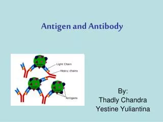
Antibody Screening and Identification. Antibody Screening. Purpose The purpose of the antibody screen is to detect red blood cell antibodies other than anti-A or anti-B. These antibodies are called “unexpected” because only 0.3 to 2 % of the general population have positive antibody screen.

E N D
Antibody Screening Purpose • The purposeof the antibody screen is to detect red blood cell antibodies other than anti-A or anti-B. • These antibodies are called “unexpected” because only 0.3 to 2 % of the general population have positive antibody screen. • Once an unexpected antibody is detected, antibody identification studies are performed to determine the antibodies specificity and clinical significance.
Antibody screening is usually undertaken at the same time as blood grouping and in advance of selecting blood for transfusion. • Antibody screening may be more reliable and sensitive than cross matching- most notably anti-Jka/Jkb but also anti-Fya, -Fyb, -S, and –s. Screening cells can be selected to reflect this, whereas donor cells are usually of unknown zygosity. • In addition, reagent red cells are easier to standardize than donor cells, and there is potentially less opportunity for procedural error, particularly in automated systems. • Clinically significant antibodies are those that are capable of causing patient morbidity as a result of accelerated destruction of a significant proportion of transfused red cells.
Antibody Screening • Antibody screening test involve testing patient’s serum against two or three reagent red blood cell samples called screening cells • Screening cells are commercially prepared group O cell suspensions obtained from individual donors that are phenotype for the most commonly encountered and clinically important red blood cell antigens. • Why group O ??
Antibody Screening • Group O cells are used so that naturally occurring anti-A or anti-B will not interfere with detection of unexpected antibodies. • The cells are selected so that the following antigens are present on at least one of the cell sample; D, C, E, c, e, M N, S, s, P, Lea, Leb, K, k, Fya, Fyb, and Jkb.
Antibody Screening • Screening Cells. • The screening cells are available in three form 1- a single vial of no more than two donors pooled together in one vial. 2- two vials each with a different donor. 3- 3 vials representing three different donors. Two or three cells screening sets are required for detection of antibodies in pre-transfusion testing.
No single test will detect all blood group Abs, but an effective compromise is as : • 1. Direct agglutination test in NISS or LISS • 2. IAT after incubation at NISS or LISS • 3. Enzyme tests; especially for detection of Rh Abs. • A positive result in Ab screen should be followed by an Ab ID against a comprehensive cell panel.
Antibody Screening • Auto-logous Control. • Autologous control is considered as part of the Ab screening, it can be performed in parallel with the Ab screen and involves testing the patient’s serum against the patient’s red blood cells. • A positive auto-logous control is an abnormal finding and usually means that patient has a positive direct antiglobulin test (DAT).
Antibody Screening • Grading Reactions. • Aggregation or hemolysis of test red blood cells is the visible end point of an Ab-Ag interaction. • Test results should be read immediately after centrifugation as delays in reading may cause elution of antibody and false- negative test results • The first step in reading hem-agglutination reactions is inspection of the supernatant for signs of hemolysis (red or pink coloration).
Why do we need to identify? • Antibody identification is needed for transfusion purposes and is an important component of compatibility testing • It will identify any unexpected antibodies in the patient’s serum • If a person with an antibody is exposed to donor cells with the corresponding antigen, serious side effects can occur .
Key Concepts • In blood banking, we test “knowns” with “unknowns” • When detecting and/or identifying antibodies, we test patient serum (unknown) with reagent RBCs (known) Known: Unknown: Reagent RBCs + patient serum Reagent antisera + patient RBCs
Reagent RBCs • Screening Cells and Panel Cells are the same with minor differences: • Screening cells • Antibody detection • Sets of 2 or 3 vials • Panel cells • Antibody identification • At least 10 vials per set
Antibody Panel vs. Screen • An antibody panel is just an extended version of an antibody screen • The screen only uses 2-3 cells:
Antibody Panel • An antibody panel usually includes at least 10 panel cells:
Panel • Group O red blood cells
Panel • Each of the panel cells has been antigen typed (shown on antigram) • + refers to the presence of the antigen • 0 refers to the absence of the antigen Example: Panel Cell #10 has 9 antigens present: c, e, f, M, s, Leb, k, Fya, and Jka
Panel • An autocontrol should also be run with ALL panels Autocontrol Patient RBCs + Patient serum
Panel • The same phases used in an antibody screen are used in a panel • IS • 37° • AHG
Antibody ID Testing • A tube is labeled for each of the panel cells plus one tube for AC: 1 2 3 4 5 6 7 8 9 10 11 AC 1 drop of each panel cell + 2 drops of the patients serum
IS Phase • Perform immediate spin (IS) and grade agglutination; inspect for hemolysis • Record the results in the appropriate space as shown: 2+ 0 0 Last tube
(LISS) 37°C Phase • 2 drops of LISS are added, mixed and incubated for 10-15 minutes • Centrifuge and check for agglutination • Record results
(LISS) 37°C Phase 2+ 0 0 0 0 0 2+ 0 0 2+ 0 2+ 0 0
IAT Phase (or AHG) • Indirect Antiglobulin Test (IAT) – we’re testing whether or not possible antibodies in patient’s serum will react with RBCs in vitro • To do this we use the Anti-Human Globulin reagent (AHG) • Polyspecific • Anti-IgG • Anti-complement
AHG Phase • Wash cells 3 times with saline (manual or automated) • Add 2 drops of AHG and gently mix • Centrifuge • Read • Record reactions
AHG Phase 2+ 0 0 0 0 0 0 0 0 2+ 0 0 0 0 0 0 0 0 2+ 0 0 0 0 0 2+ 0 0 0 0 0 0 0 0 0 0 0
And don’t forget…. ….add “check” cells to any negative AHG !
You have agglutination…now what? CC 2+ 0 0 0 0 0 0 0 0 2+ 0 0 0 0 0 0 0 0 2+ 0 0 0 0 0 2+ 0 0 0 0 0 0 0 0 0 0 0 ??
Interpreting Antibody Panels • There are a few basic steps to follow when interpreting panels • “Ruling out” means crossing out antigens that did not react • Circle the antigens that are not crossed out • Consider antibody’s usual reactivity • Look for a matching pattern
Always remember: An antibody will only react with cells that have the corresponding antigen; antibodies will not react with cells that do not have the antigen
1. Ruling Out 2+ 0 0 0 0 0 0 0 0 2+ 0 0 0 0 0 0 0 0 2+ 0 0 0 0 0 2+ 0 0 0 0 0 0 0 0 0 0 0 Cross out antigens that show NO REACTION in any phase; do NOTcross out heterozygous antigens that show dosage.
About reaction strengths…… • Strength of reaction may be due to “dosage” • If panel cells are homozygous, a strong reaction may be seen • If panel cells are heterozygous, reaction may be weak or even non-reactive • Panel cells that are heterozygous should not be crossed out because antibody may be too weak to react
2. Circle antigens not crossed out 2+ 0 0 0 0 0 0 0 0 2+ 0 0 0 0 0 0 0 0 2+ 0 0 0 0 0 2+ 0 0 0 0 0 0 0 0 0 0 0
3. Consider antibody’s usual reactivity 2+ 0 0 0 0 0 0 0 0 2+ 0 0 0 0 0 0 0 0 2+ 0 0 0 0 0 2+ 0 0 0 0 0 0 0 0 0 0 0 Lea is normally a Cold-Reacting antibody (IgM), so it makes sense that we see the reaction in the IS phase of testing; The E antigen will usually react at warmer temperatures
4. Look for a matching pattern E doesn’t match and it’s a warmer rx Ab 2+ 0 0 0 0 0 0 0 0 2+ 0 0 0 0 0 0 0 0 2+ 0 0 0 0 0 2+ 0 0 0 0 0 0 0 0 0 0 0 …Yes, there is a matching pattern!
Interpretation anti-Lea
Guidelines • Again, it’s important to look at: • Autocontrol • Negative - alloantibody • Positive – autoantibody or DTR (i.e.,alloantibodies) • Phases • IS – cold (IgM) • 37° - cold (some have higher thermal range) or warm reacting • AHG – warm (IgG)…significant!! • Reaction strength • 1 consistent strength – one antibody • Different strengths – multiple antibodies or dosage
Guidelines (continued) • Matching the pattern • Single antibodies usually shows a pattern that matches one of the antigens (see previous panel example) • Multiple antibodies are more difficult to match because they often show mixed reaction strengths
Rule of three • The rule of three must be met to confirm the presence of the antibody • How is it demonstrated? • Patient serum MUST be: • Positive with 3 cells with the antigen • Negative with 3 cells without the antigen
Our previous example fulfills the “rule of three” 2+ 0 0 0 0 0 3 Positive cells 0 0 0 2+ 0 0 0 0 0 0 0 0 2+ 0 0 0 0 0 3 Negative cells 2+ 0 0 0 0 0 0 0 0 0 0 0 Panel Cells 1, 4, and 7 are positive for the antigen and gave a reaction at immediate spin Panel Cells 8, 10, and 11 are negative for the antigen and did not give a reaction at immediate spin
What if the “rule of three” is not fulfilled? • If there are not enough cells in the panel to fulfill the rule, then additional cells from another panel could be used • Most labs carry different lot numbers of panel cells