Download
1 / 32
700 likes | 1.63k Views
© 2012 Pearson Education, Inc. I. Cell Shape and Size. 3.1 Cell Morphology 3.2 Cell Size and the Significance of Smallness. Figure 3.1. Spirochete. Coccus. Hypha. Stalk. Rod. Budding and appendaged bacteria. Spirillum. Filamentous bacteria. © 2012 Pearson Education, Inc.

E N D
© 2012 Pearson Education, Inc. I. Cell Shape and Size • 3.1 Cell Morphology • 3.2 Cell Size and the Significance of Smallness
Figure 3.1 Spirochete Coccus Hypha Stalk Rod Budding and appendaged bacteria Spirillum Filamentous bacteria © 2012 Pearson Education, Inc.
© 2012 Pearson Education, Inc. 3.1 Cell Morphology • Morphology typically does not predict physiology, ecology, phylogeny, etc. of a prokaryotic cell • Selective forces may be involved in setting the morphology • Optimization for nutrient uptake (small cells and those with high surface-to-volume ratio) • Swimming motility in viscous environments or near surfaces (helical or spiral-shaped cells) • Gliding motility (filamentous bacteria)
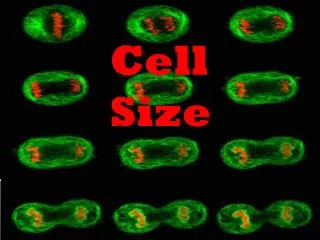
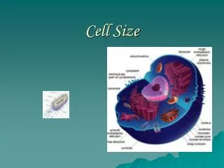
© 2012 Pearson Education, Inc. 3.2 Cell Size and the Significance of Smallness • Size range for prokaryotes: 0.2 µm to >700 µm in diameter • Most cultured rod-shaped bacteria are between 0.5 and 4.0 µm wide and <15 µm long • Examples of very large prokaryotes • Epulopiscium fishelsoni (Figure 3.2a) • Thiomargarita namibiensis (Figure 3.2b) • Size range for eukaryotic cells: 10 to >200 µm in diameter
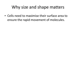
© 2012 Pearson Education, Inc. 3.2 Cell Size and the Significance of Smallness • Surface-to-Volume Ratios, Growth Rates, and Evolution • Advantages to being small (Figure 3.3) • Small cells have more surface area relative to cell volume than large cells (i.e., higher S/V) • support greater nutrient exchange per unit cell volume • tend to grow faster than larger cells
© 2012 Pearson Education, Inc. 3.3 The Cytoplasmic Membrane in Bacteria and Archaea • Cytoplasmic membrane: • Thin structure that surrounds the cell • 6–8 nm thick • Vital barrier that separates cytoplasm from environment • Highly selective permeable barrier; enables concentration of specific metabolites and excretion of waste products
© 2012 Pearson Education, Inc. 3.3 The Cytoplasmic Membrane • Composition of Membranes • General structure is phospholipid bilayer (Figure 3.4) • Contain both hydrophobic and hydrophilic components • Can exist in many different chemical forms as a result of variation in the groups attached to the glycerol backbone • Fatty acids point inward to form hydrophobic environment; hydrophilic portions remain exposed to external environment or the cytoplasm Animation: Membrane Structure
Figure 3.4 Glycerol Fatty acids Phosphate Ethanolamine Hydrophilic region Hydrophobic Fatty acids region Hydrophilic region Glycerophosphates Fatty acids © 2012 Pearson Education, Inc.
© 2012 Pearson Education, Inc. 3.3 The Cytoplasmic Membrane • Cytoplasmic Membrane (Figure 3.5) • 6–8 nm wide • Embedded proteins • Stabilized by hydrogen bonds and hydrophobic interactions • Mg2+ and Ca2+ help stabilize membrane by forming ionic bonds with negative charges on the phospholipids • Somewhat fluid
Figure 3.5 Out Phospholipids Hydrophilic groups 6–8 nm Hydrophobic groups In Integral Phospholipid membrane proteins molecule © 2012 Pearson Education, Inc.
Figure 3.6 Ester Ether Bacteria Archaea Eukarya © 2012 Pearson Education, Inc.
Figure 3.7d Out Glycerophosphates Phytanyl Membrane protein In Lipid bilayer © 2012 Pearson Education, Inc.
Figure 3.7e Out Biphytanyl In Lipid monolayer © 2012 Pearson Education, Inc.
© 2012 Pearson Education, Inc. 3.4 Functions of the Cytoplasmic Membrane • Permeability Barrier (Figure 3.8) • Polar and charged molecules must be transported • Transport proteins accumulate solutes against the concentration gradient • Protein Anchor • Holds transport proteins in place • Energy Conservation
© 2012 Pearson Education, Inc. III. Cell Walls of Prokaryotes • 3.6 The Cell Wall of Bacteria: Peptidoglycan • 3.7 The Outer Membrane • 3.8 Cell Walls of Archaea
© 2012 Pearson Education, Inc. 3.6 The Cell Wall of Bacteria: Peptidoglycan Peptidoglycan (Figure 3.16) • Rigid layer that provides strength to cell wall • Polysaccharide composed of • N-acetylglucosamine and N-acetylmuramic acid • Amino acids • Lysine or diaminopimelic acid (DAP) • Cross-linked differently in gram-negative bacteria and gram-positive bacteria (Figure 3.17)
Figure 3.17 Polysaccharide backbone Interbridge Peptides Escherichia coli (gram-negative) Staphylococcus aureus (gram-positive) Y Peptide bonds X Glycosidic bonds © 2012 Pearson Education, Inc.
© 2012 Pearson Education, Inc. 3.6 The Cell Wall of Bacteria: Peptidoglycan • Gram-Positive Cell Walls (Figure 3.18) • Can contain up to 90% peptidoglycan • Common to have teichoic acids (acidic substances) embedded in the cell wall • Lipoteichoic acids: teichoic acids covalently bound to membrane lipids
Figure 3.18 Peptidoglycan cable Ribitol Teichoic acid Peptidoglycan Wall-associated protein Lipoteichoic acid Cytoplasmic membrane © 2012 Pearson Education, Inc.
© 2012 Pearson Education, Inc. 3.6 The Cell Wall of Bacteria: Peptidoglycan • Prokaryotes That Lack Cell Walls • Mycoplasmas • Group of pathogenic bacteria • Thermoplasma • Species of Archaea
© 2012 Pearson Education, Inc. 3.7 The Outer Membrane • Total cell wall contains ~10% peptidoglycan (Figure 3.20a) • Most of cell wall composed of outer membrane (aka lipopolysaccharide [LPS] layer) • LPS consists of core polysaccharide and O-polysaccharide • LPS replaces most of phospholipids in outer half of outer membrane • Endotoxin: the toxic component of LPS
Figure 3.20a O-polysaccharide Core polysaccharide Out Lipid A Protein Lipopolysaccharide (LPS) Porin 8 nm Outer membrane Cell wall Phospholipid Peptidoglycan Periplasm Lipoprotein Cytoplasmic membrane In © 2012 Pearson Education, Inc.
© 2012 Pearson Education, Inc. 3.7 The Outer Membrane • Structural differences between cell walls of gram-positive and gram-negative Bacteria are responsible for differences in the Gram stain reaction
© 2012 Pearson Education, Inc. 3.8 Cell Walls of Archaea • No peptidoglycan • Typically no outer membrane • Pseudomurein • Polysaccharide similar to peptidoglycan (Figure 3.21) • Composed of N-acetylglucosamine and N-acetyltalosaminuronic acid • Found in cell walls of certain methanogenic Archaea • Cell walls of some Archaea lack pseudomurein
© 2012 Pearson Education, Inc. 3.8 Cell Walls of Archaea • S-Layers • Most common cell wall type among Archaea • Consist of protein or glycoprotein • Paracrystalline structure (Figure 3.22)
Figure 3.22 © 2012 Pearson Education, Inc.
© 2012 Pearson Education, Inc. 3.9 Cell Surface Structures • Capsules and Slime Layers • Polysaccharide layers (Figure 3.23) • May be thick or thin, rigid or flexible • Assist in attachment to surfaces • Protect against phagocytosis • Resist desiccation
Figure 3.23 Cell Capsule © 2012 Pearson Education, Inc.
© 2012 Pearson Education, Inc. 3.9 Cell Surface Structures • Fimbriae • Filamentous protein structures (Figure 3.24) • Enable organisms to stick to surfaces or form pellicles
Figure 3.24 Flagella Fimbriae © 2012 Pearson Education, Inc.
© 2012 Pearson Education, Inc. 3.9 Cell Surface Structures • Pili • Filamentous protein structures (Figure 3.25) • Typically longer than fimbriae • Assist in surface attachment • Facilitate genetic exchange between cells (conjugation) • Type IV pili involved in twitching motility
Figure 3.25 Virus- covered pilus © 2012 Pearson Education, Inc.