Download
1 / 64
640 likes | 716 Views
16. The Endocrine System: Part B. The Posterior Pituitary. Contains axons of hypothalamic neurons Stores antidiuretic hormone (ADH) and oxytocin ADH and oxytocin are released in response to nerve impulses Both use PIP-calcium second-messenger mechanism at their targets. Oxytocin.

E N D
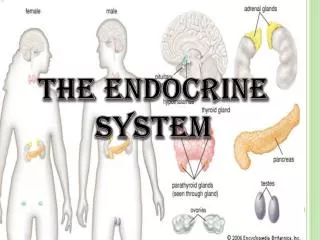
16 The Endocrine System: Part B
The Posterior Pituitary • Contains axons of hypothalamic neurons • Stores antidiuretic hormone (ADH) and oxytocin • ADH and oxytocin are released in response to nerve impulses • Both use PIP-calcium second-messenger mechanism at their targets
Oxytocin • Stimulates uterine contractions during childbirth by mobilizing Ca2+ through a PIP2-Ca2+ second-messenger system • Also triggers milk ejection (“letdown” reflex) in women producing milk • Plays a role in sexual arousal and orgasm in males and females
Antidiuretic Hormone (ADH) • Hypothalamic osmoreceptors respond to changes in the solute concentration of the blood • If solute concentration is high • Osmoreceptors depolarize and transmit impulses to hypothalamic neurons • ADH is synthesized and released, inhibiting urine formation
Antidiuretic Hormone (ADH) • If solute concentration is low • ADH is not released, allowing water loss • Alcohol inhibits ADH release and causes copious urine output
Homeostatic Imbalances of ADH • ADH deficiency—diabetes insipidus; huge output of urine and intense thirst • ADH hypersecretion (after neurosurgery, trauma, or secreted by cancer cells)—syndrome of inappropriate ADH secretion (SIADH)
Thyroid Gland • Consists of two lateral lobes connected by a median mass called the isthmus • Composed of follicles that produce the glycoprotein thyroglobulin • Colloid (thyroglobulin + iodine) fills the lumen of the follicles and is the precursor of thyroid hormone • Parafollicular cells produce the hormone calcitonin
Thyroid Hormone (TH) • Actually two related compounds • T4 (thyroxine); has 2 tyrosine molecules + 4 bound iodine atoms • T3 (triiodothyronine); has 2 tyrosines + 3 bound iodine atoms
Thyroid Hormone • Major metabolic hormone • Increases metabolic rate and heat production (calorigenic effect) • Plays a role in • Maintenance of blood pressure • Regulation of tissue growth • Development of skeletal and nervous systems • Reproductive capabilities
Synthesis of Thyroid Hormone • Thyroglobulin is synthesized and discharged into the follicle lumen • Iodides (I–) are actively taken into the cell, oxidized to iodine (I2), and released into the lumen • Iodine attaches to tyrosine, mediated by peroxidase enzymes
Synthesis of Thyroid Hormone • Iodinated tyrosines link together to form T3 and T4 • Colloid is endocytosed and combined with a lysosome • T3 and T4 are cleaved and diffuse into the bloodstream
Thyroid follicle cells Colloid 1 Thyroglobulin is synthesized anddischarged into the follicle lumen. Tyrosines (part of thyroglobulinmolecule) Capillary 4 Iodine is attached to tyrosinein colloid, forming DIT and MIT. Golgiapparatus RoughER Thyro-globulincolloid Iodine DIT (T2) MIT (T1) 3 Iodideis oxidizedto iodine. 2 Iodide (I–) is trapped(actively transported in). Iodide (I–) T4 5 Iodinated tyrosines arelinked together to form T3and T4. T3 Lysosome T4 6 Thyroglobulin colloid isendocytosed and combinedwith a lysosome. T3 7 Lysosomal enzymes cleaveT4 and T3 from thyroglobulincolloid and hormones diffuseinto bloodstream. Colloid inlumen offollicle T4 T3 To peripheral tissues Figure 16.9
Thyroid follicle cells Colloid 1 Thyroglobulin is synthesized anddischarged into the follicle lumen. Tyrosines (part of thyroglobulinmolecule) Capillary Golgiapparatus RoughER Colloid inlumen offollicle Figure 16.9, step 1
Thyroid follicle cells Colloid 1 Thyroglobulin is synthesized anddischarged into the follicle lumen. Tyrosines (part of thyroglobulinmolecule) Capillary Golgiapparatus RoughER 2 Iodide (I–) is trapped(actively transported in). Iodide (I–) Colloid inlumen offollicle Figure 16.9, step 2
Thyroid follicle cells Colloid 1 Thyroglobulin is synthesized anddischarged into the follicle lumen. Tyrosines (part of thyroglobulinmolecule) Capillary Golgiapparatus RoughER Iodine 3 Iodideis oxidizedto iodine. 2 Iodide (I–) is trapped(actively transported in). Iodide (I–) Colloid inlumen offollicle Figure 16.9, step 3
Thyroid follicle cells Colloid 1 Thyroglobulin is synthesized anddischarged into the follicle lumen. Tyrosines (part of thyroglobulinmolecule) Capillary 4 Iodine is attached to tyrosinein colloid, forming DIT and MIT. Golgiapparatus RoughER Thyro-globulincolloid Iodine DIT (T2) MIT (T1) 3 Iodideis oxidizedto iodine. 2 Iodide (I–) is trapped(actively transported in). Iodide (I–) Colloid inlumen offollicle Figure 16.9, step 4
Thyroid follicle cells Colloid 1 Thyroglobulin is synthesized anddischarged into the follicle lumen. Tyrosines (part of thyroglobulinmolecule) Capillary 4 Iodine is attached to tyrosinein colloid, forming DIT and MIT. Golgiapparatus RoughER Thyro-globulincolloid Iodine DIT (T2) MIT (T1) 3 Iodideis oxidizedto iodine. 2 Iodide (I–) is trapped(actively transported in). Iodide (I–) T4 5 Iodinated tyrosines arelinked together to form T3and T4. T3 Colloid inlumen offollicle Figure 16.9, step 5
Thyroid follicle cells Colloid 1 Thyroglobulin is synthesized anddischarged into the follicle lumen. Tyrosines (part of thyroglobulinmolecule) Capillary 4 Iodine is attached to tyrosinein colloid, forming DIT and MIT. Golgiapparatus RoughER Thyro-globulincolloid Iodine DIT (T2) MIT (T1) 3 Iodideis oxidizedto iodine. 2 Iodide (I–) is trapped(actively transported in). Iodide (I–) T4 5 Iodinated tyrosines arelinked together to form T3and T4. T3 Lysosome 6 Thyroglobulin colloid isendocytosed and combinedwith a lysosome. Colloid inlumen offollicle Figure 16.9, step 6
Thyroid follicle cells Colloid 1 Thyroglobulin is synthesized anddischarged into the follicle lumen. Tyrosines (part of thyroglobulinmolecule) Capillary 4 Iodine is attached to tyrosinein colloid, forming DIT and MIT. Golgiapparatus RoughER Thyro-globulincolloid Iodine DIT (T2) MIT (T1) 3 Iodideis oxidizedto iodine. 2 Iodide (I–) is trapped(actively transported in). Iodide (I–) T4 5 Iodinated tyrosines arelinked together to form T3and T4. T3 Lysosome T4 6 Thyroglobulin colloid isendocytosed and combinedwith a lysosome. T3 7 Lysosomal enzymes cleaveT4 and T3 from thyroglobulincolloid and hormones diffuseinto bloodstream. Colloid inlumen offollicle T4 T3 To peripheral tissues Figure 16.9, step 7
Transport and Regulation of TH • T4 and T3 are transported by thyroxine-binding globulins (TBGs) • Both bind to target receptors, but T3 is ten times more active than T4 • Peripheral tissues convert T4 to T3
Transport and Regulation of TH • Negative feedback regulation of TH release • Rising TH levels provide negative feedback inhibition on release of TSH • Hypothalamic thyrotropin-releasing hormone (TRH) can overcome the negative feedback during pregnancy or exposure to cold
Hypothalamus TRH Anterior pituitary TSH Thyroid gland Thyroid hormones Stimulates Target cells Inhibits Figure 16.7
Homeostatic Imbalances of TH • Hyposecretion in adults—myxedema; endemic goiter if due to lack of iodine • Hyposecretion in infants—cretinism • Hypersecretion—Graves’ disease
Calcitonin • Produced by parafollicular (C) cells • Antagonist to parathyroid hormone (PTH) • Inhibits osteoclast activity and release of Ca2+ from bone matrix
Calcitonin • Stimulates Ca2+ uptake and incorporation into bone matrix • Regulated by a humoral (Ca2+ concentration in the blood) negative feedback mechanism • No important role in humans; removal of thyroid (and its C cells) does not affect Ca2+ homeostasis
Parathyroid Glands • Four to eight tiny glands embedded in the posterior aspect of the thyroid • Contain oxyphil cells (function unknown) and chief cells that secrete parathyroid hormone (PTH) or parathormone • PTH—most important hormone in Ca2+ homeostasis
Pharynx (posterior aspect) Chief cells (secrete parathyroid hormone) Thyroid gland Parathyroid glands Oxyphil cells Esophagus Trachea Capillary (a) (b) Figure 16.11
Parathyroid Hormone • Functions • Stimulates osteoclasts to digest bone matrix • Enhances reabsorption of Ca2+ and secretion of phosphate by the kidneys • Promotes activation of vitamin D (by the kidneys); increases absorption of Ca2+ by intestinal mucosa • Negative feedback control: rising Ca2+ in the blood inhibits PTH release
Hypocalcemia (low blood Ca2+) stimulates parathyroid glands to release PTH. Rising Ca2+ in blood inhibits PTH release. Bone 1 PTH activates osteoclasts: Ca2+ and PO43Sreleased into blood. Kidney 2 PTH increases Ca2+reabsorption in kidney tubules. 3 PTH promotes kidney’s activation of vitamin D, which increases Ca2+ absorption from food. Intestine Ca2+ ions Bloodstream PTH Molecules Figure 16.12
Homeostatic Imbalances of PTH • Hyperparathyroidism due to tumor • Bones soften and deform • Elevated Ca2+ depresses the nervous system and contributes to formation of kidney stones • Hypoparathyroidism following gland trauma or removal • Results in tetany, respiratory paralysis, and death
Adrenal (Suprarenal) Glands • Paired, pyramid-shaped organs atop the kidneys • Structurally and functionally, they are two glands in one • Adrenal medulla—nervous tissue; part of the sympathetic nervous system • Adrenal cortex—three layers of glandular tissue that synthesize and secrete corticosteroids
Adrenal Cortex • Three layers and the corticosteroids produced • Zona glomerulosa—mineralocorticoids • Zona fasciculata—glucocorticoids • Zona reticularis—sex hormones, or gonadocorticoids
Capsule Zona glomerulosa Zona fasciculata Adrenal gland Cortex • Medulla • Cortex Zona reticularis Kidney Adrenal medulla Medulla (a) Drawing of the histology of the adrenal cortex and a portion of the adrenal medulla Figure 16.13a
Mineralocorticoids • Regulate electrolytes (primarily Na+ and K+) in ECF • Importance of Na+: affects ECF volume, blood volume, blood pressure, levels of other ions • Importance of K+: sets RMP of cells • Aldosterone is the most potent mineralocorticoid • Stimulates Na+ reabsorption and water retention by the kidneys
Mechanisms of Aldosterone Secretion • Renin-angiotensin mechanism: decreased blood pressure stimulates kidneys to release renin, triggers formation of angiotensin II, a potent stimulator of aldosterone release • Plasma concentration of K+: Increased K+ directly influences the zona glomerulosa cells to release aldosterone • ACTH: causes small increases of aldosterone during stress • Atrial natriuretic peptide (ANP): blocks renin and aldosterone secretion, to decrease blood pressure
Primary regulators Other factors Blood volume and/or blood pressure K+in blood Stress Blood pressure and/or blood volume Hypo- thalamus Heart Kidney CRH Direct stimulating effect Anterior pituitary Renin Initiates cascade that produces Atrial natriuretic peptide (ANP) ACTH Angiotensin II Inhibitory effect Zona glomerulosa of adrenal cortex Enhanced secretion of aldosterone Targets kidney tubules Absorption of Na+ and water; increased K+ excretion Blood volume and/or blood pressure Figure 16.14
Homeostatic Imbalances of Aldosterone • Aldosteronism—hypersecretion due to adrenal tumors • Hypertension and edema due to excessive Na+ • Excretion of K+ leading to abnormal function of neurons and muscle
Glucocorticoids (Cortisol) • Keep blood sugar levels relatively constant • Maintain blood pressure by increasing the action of vasoconstrictors
Glucocorticoids (Cortisol) • Cortisol is the most significant glucocorticoid • Released in response to ACTH, patterns of eating and activity, and stress • Prime metabolic effect is gluconeogenesis—formation of glucose from fats and proteins • Promotes rises in blood glucose, fatty acids, and amino acids
Homeostatic Imbalances of Glucocorticoids • Hypersecretion—Cushing’s syndrome • Depresses cartilage and bone formation • Inhibits inflammation • Depresses the immune system • Promotes changes in cardiovascular, neural, and gastrointestinal function • Hyposecretion—Addison’s disease • Also involves deficits in mineralocorticoids • Decrease in glucose and Na+ levels • Weight loss, severe dehydration, and hypotension
Gonadocorticoids (Sex Hormones) • Most are androgens (male sex hormones) that are converted to testosterone in tissue cells or estrogens in females • May contribute to • The onset of puberty • The appearance of secondary sex characteristics • Sex drive
Adrenal Medulla • Chromaffin cells secrete epinephrine (80%) and norepinephrine (20%) • These hormones cause • Blood glucose levels to rise • Blood vessels to constrict • The heart to beat faster • Blood to be diverted to the brain, heart, and skeletal muscle
Adrenal Medulla • Epinephrine stimulates metabolic activities, bronchial dilation, and blood flow to skeletal muscles and the heart • Norepinephrine influences peripheral vasoconstriction and blood pressure
Short-term stress More prolonged stress Stress Nerve impulses Hypothalamus CRH (corticotropin- releasing hormone) Spinal cord Corticotroph cells of anterior pituitary Preganglionic sympathetic fibers To target in blood Adrenal cortex (secretes steroid hormones) Adrenal medulla (secretes amino acid- based hormones) ACTH Catecholamines (epinephrine and norepinephrine) Mineralocorticoids Glucocorticoids Short-term stress response Long-term stress response 1. Increased heart rate 2. Increased blood pressure 3. Liver converts glycogen to glucose and releases glucose to blood 4. Dilation of bronchioles 5. Changes in blood flow patterns leading to decreased digestive system activity and reduced urine output 6. Increased metabolic rate 1. Retention of sodium and water by kidneys 2. Increased blood volume and blood pressure 1. Proteins and fats converted to glucose or broken down for energy 2. Increased blood glucose 3. Suppression of immune system Figure 16.16
Pineal Gland • Small gland hanging from the roof of the third ventricle • Pinealocytes secrete melatonin, derived from serotonin • Melatonin may affect • Timing of sexual maturation and puberty • Day/night cycles • Physiological processes that show rhythmic variations (body temperature, sleep, appetite)
Pancreas • Triangular gland behind the stomach • Has both exocrine and endocrine cells • Acinar cells (exocrine) produce an enzyme-rich juice for digestion • Pancreatic islets (islets of Langerhans) contain endocrine cells • Alpha () cells produce glucagon (a hyperglycemic hormone) • Beta () cells produce insulin (a hypoglycemic hormone)
Pancreatic islet (of Langerhans) • (Glucagon- producing) cells • (Insulin- producing) cells Pancreatic acinar cells (exocrine) Figure 16.17