Download
1 / 136
1.48k likes | 1.99k Views
The Human Body. By Albert Wu. Organ Systems. Digestive System Circulatory System Respiratory System Excretory System Immune System. Skeletal System Endocrine System Sensory System Muscular System Nervous System Reproductive System. The Digestive System. Functions.

E N D
The Human Body By Albert Wu
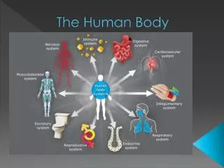
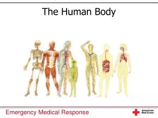
Organ Systems • Digestive System • Circulatory System • Respiratory System • Excretory System • Immune System • Skeletal System • Endocrine System • Sensory System • Muscular System • Nervous System • Reproductive System
Functions • Provides intake of food for nourishment • Breaks down the food into components which can be absorbed and utilized by the body • Excretes solid waste
The Process of Digestion • The digestive tract absorbs food mainly by diffusion or active transport. • Large food molecules can’t diffuse and are too large to fit the active transport pumps. • Therefore, food molecules have to be broken down and absorbed. The digestive system performs both of these functions.
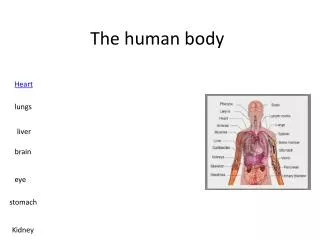
Parts of the Digestive System Teeth Salivary Glands Lip Mouth Tongue Pharynx Esophagus Liver Stomach Gallbladder (under liver) Pancreas (under stomach) Large Intestine Rectum Small Intestine Anus Appendix
The Alimentary Canal • The alimentary canal consists of the organs that form the main digestive tract. • The alimentary canal includes: • Mouth, Pharynx, and Esophagus • Stomach • Small Intestine • Large Intestine • Rectum and Anus
The Mouth, Pharynx, and Esophagus • The mouth physically breaks down food (using the teeth) and also begins to chemically digest carbohydrates. The tongue senses different foods. • The pharynx connects the mouth to the esophagus, which pushes food into the stomach by means of peristaltic contractions. The food enters the stomach via the esophageal sphincter. The mouth and associated organs.
The Stomach • The stomach is a muscular pouch that receives chewed food from the esophagus. • The stomach secretes hydrochloric acid as well as a number of enzymes which chemically digest proteins in food. The half-digested food is called chyme. • Muscular contractions of the stomach wall help mix the digesting food and push it into the small intestine through a valve called the pyloric sphincter.
The Small Intestine • The final stages of chemical breakdown of food occur in the small intestine, which is a long muscular tube. • Most nutrients are absorbed in the small intestine. • The small intestine’s great length (5.5-6 meters) and internal folds (villi and microvilli) helps give the maximum surface area for absorption of food. • Muscular contractions mix the food and move it down the intestine. Right: the lining of the small intestine, seen through a microscope.
Parts of the Small Intestine • The small intestine has three main sections. In order from the closest to the stomach, they are the duodenum, the jejunum, and the ileum. • The first 25 cm is the duodenum. Pancreatic and liver secretions enter the duodenum. • The next 2.5 m is the jejunum. It is relatively thick and exhibits high activity in its vascular wall. The pH of the jejunum and the ileum ranges from 7-9. • The final 2-4 m is the ileum. The ileum houses a large population of symbiotic bacteria and has many lymph nodes.
The Large Intestine • The chyme (now mostly digested) enters the large intestine through the ileocecal sphincter. The large intestine absorbs water and electrolytes from it. • Indigestible waste material (feces) is pushed into the rectum. • The large intestine is shorter than the small intestine but much wider.
Parts of the Large Intestine. • The large intestine has five main parts. They occur in this order, beginning with the closest to the small intestine. • The cecum is a pouch that hangs below the ileocecal sphincter. It is connected to the appendix, a small, wormlike structure. • The ascending colon stretches up from the cecum to the liver. • The transverse colon extends across the abdomen. • The descending colon drops down to the pelvic girdle. • The sigmoid colon is s-shaped and connects to the rectum.
The Rectum and Anus • The rectum collects feces (indigestible material) and periodically excretes them (defecation). • The anus is a muscular ring that acts as a valve for the rectum.
Accessory Organs • The accessory organs secrete substances into the alimentary canal which assist the process of digestion. They are not actually part of the alimentary canal. • The accessory organs include: • Salivary Glands • Pancreas • Liver and Gallbladder
The Salivary Glands • The salivary glands produce saliva, which contains enzymes that break down carbohydrates and kill certain pathogens. • Saliva also contains bicarbonate ions which buffer the saliva to prevent enzyme denaturation.
The Pancreas • The pancreas produces “pancreatic juice”, which is secreted into the small intestine. • Pancreatic juice contains a variety of enzymes, including those listed below. • Amylase: digests starch • Lipase: digests lipids • Trypsin: digests proteins • Nuclease: digests nucleic acids • Pancreatic juice also contains bicarbonate ions, which make an optimum environment for pancreatic enzymes and help neutralize stomach acid. • The pancreas is also part of the endocrine system. The pancreas.
The Liver and Gallbladder • The liver and gallbladder help digest lipids. • The liver secretes a liquid called bile, which contains pigments, salts, and cholesterol. • The gallbladder collects bile, concentrates it, and secretes it into the small intestine. • Bile salts cause lipids to form globules (emulsification) and aid enzymes in digesting the lipids.
Enzymes • Enzymes are proteins which act as catalysts. • They are important in breaking down large food molecules. • Different parts of the digestive system produce different enzymes. • Digestion of different types of food molecules takes place in different locations.
Physical and Chemical Digestion Physical Digestion Chemical Digestion The physical breakdown of pieces of food Increases surface area of food for chemical digestion to be effective The chemical breakdown of food molecules Breaks large molecules into smaller molecules which can be absorbed Happens at a molecular level
Diseases of the Digestive System • Pinworms • Caused by a small parasitic worm, Enterobiusvermicularis. • Symptoms: itching in the crotch area, may cause weight loss/anemia. • Nearly 1/3 of the world’s population is infected with pinworms, but symptoms are usually mild. • Treated with repeated doses of medication to ensure the death of all the pinworm eggs.
Diseases (continued) • Colorectal Cancer • Caused by uncontrolled mitosis of cells in the large intestine • Symptoms include abdominal pain, bloody stools or diarrhea, or changes in bowel movements. • 4th most common cancer in world, more common in older people or alcoholics, 20% have family history • Treated with radiation or chemotherapy
Sources • Shier, David, Jackie Butler, and Ricki Lewis. Hole’s Human Anatomy and Physiology. New York, McGraw-Hill: 2007. • http://www.google.com/ • http://en.wikipedia.org/ • http://www.ncbi.nlm.nih.gov/ • http://digestive.niddk.nih.gov/ddiseases/pubs/yrdd/
To move blood around the body, ensuring that all tissues get an adequate supply of nutrients and oxygen. Wastes and carbon dioxide are removed. • To circulate white blood cells for defense against infection and to circulate platelets for damage repair. Functions
The heart is a muscular pump that contracts forcefully to pump blood through the blood vessels. • The heart has four chambers to pump blood in two circuits: the pulmonary circuit (to the lungs) and the systemic circuit (to the body). • The heart’s rate of contraction can change to suit the body’s oxygen or nutrition needs (example: heart rate increases during exercise). The Heart
The Heart Aorta Superior Vena Cava Left Pulmonary Artery Right Pulmonary Artery Left Pulmonary Veins Right Pulmonary Veins Left Atrium Pulmonary Trunk Right Atrium Mitral Valve Coronary Vessels Aortic Valve Tricuspid Valve Left Ventricle Pulmonary Valve Chordae Tendineae Right Ventricle Papillary Muscles Interventricular Septum Inferior Vena Cava
1. Deoxygenated (oxygen poor) blood arrives at the right atrium through the venae cavae, the right atrium contracts, the blood moves to right ventricle through tricuspid valve, the tricuspid valve closes. • 2. The right ventricle contracts, pumping blood through the pulmonary valve and pulmonary artery to lungs where the blood picks up oxygen, the pulmonary valve closes. • 3. Oxygenated blood arrives at left atrium via pulmonary veins, left atrium contracts, the blood moves to left ventricle through mitral valve, the mitral valve closes. How the Heart Works See the heart diagram on slide 25.
4. Left ventricle contracts, pumping oxygenated blood through the aortic valve and aorta into the body, where the blood gives up its oxygen. The cycle starts again. • The tricuspid and mitral valves are operated by the chordae tendineae and the papillary muscles. The pulmonary and aortic valves are semilunar valves, which are closed by blood pressure. How the Heart Works
There are several types of blood vessels. • Arteries and arterioles: carry blood away from the heart. • Capillaries: allow exchange of oxygen and nutrients between blood and tissues. • Veins and venules: carry blood back to the heart. Here is a cross section of an artery (right) and a vein (left). Notice the difference in thickness of the walls of the vessels. Blood Vessels
Arteries and arterioles carry blood away from the heart. Arterioles are small arteries. • Blood pumped from the heart is under high pressure, so arteries and arterioles have a very thick and muscular wall to deal with the pressure. Arteries and Arterioles A cross section of an artery.
Veins and venules carry blood back to the heart. • Veins and venules have a wide diameter to carry large volumes of blood. • Veins and venules have thin walls because the blood is at low pressures. • Low pressure blood tends to flow back down the veins due to gravity, so veins are equipped with internal valves. Veins and Venules A cross section of a vein.
Capillaries are the smallest blood vessels – they are so small that red blood cells can only pass through single file. • The thin walls of capillaries (only one cell thick) facilitate the exchange of nutrients and waste, along with oxygen and carbon dioxide. Capillaries
Blood is a connective tissue and the only liquid tissue in the body. It is pumped through the blood vessels and lungs by the heart. • Blood is important for homeostasis. It carries oxygen and nutrients to tissues and takes away carbon dioxide and wastes. • White blood cells, which fight infections, are circulated through the body through the blood. The Blood
Blood is composed of plasma, red blood cells, white blood cells, and platelets. • 55% of blood is composed of plasma, a watery fluid which contains proteins important for homeostasis and blood clotting. Composition of Blood
Nearly 45% of blood is made up of red blood cells. • Red blood cells carry oxygen and carbon dioxide. They have an iron-containing protein called hemoglobin, which can bind to oxygen and carbon dioxide. • Red blood cells have a biconcave disc shape which allows them to squeeze through narrow capillaries to perform oxygen exchange. Red Blood Cells Notice the unique shape of these red blood cells.
White blood cells fight infections. There are two main types: granulocytes and agranulocytes. • Granulocytes • Neutrophil: phagocytize bacteria and small particles • Eosinophil: kill parasites and regulate allergic responses • Basophil: regulate immune response by secreting histamine • Agranulocytes • Monocyte: differentiate into macrophages (phagocytize large particles) • Lymphocytes • T lymphocytes: kill virus-infected cells and cancers • B lymphocytes: produce antibodies White Blood Cells
Platelets are small cell fragments that are produced in the bone marrow. They are important in homeostasis and healing of wounds. • If a blood vessel is punctured, platelets form a plug to prevent blood loss. They also secrete proteins which combine with other blood proteins to form a sticky mesh. This mesh entangles other cells to create a clot. Platelets
There are two types of circulatory systems: open and closed. • A closed circulatory system has blood contained within vessels. Most vertebrates (including humans) have a closed circulatory system. • An open circulatory system simply circulates blood within the main body cavity. Most insects and invertebrates have an open circulatory system. Open and Closed Circulatory Systems A fish (on the left) has a closed circulatory system. A crayfish (right) has an open circulatory system.
Not all animals have hearts similar to those of humans. • Mammals (including humans) and birds have a complete four-chambered heart (2 atria, 2 ventricles). • Amphibians have a three-chambered heart (2 atria, 1 ventricle) with channels that reduce mixing of blood between the two circuits. Reptile hearts are similar, but the ventricle has an incomplete partition. • Fish have a two-chambered heart (1 atrium, 1 ventricle) that only pumps blood in one circuit. Hearts of Vertebrates
Circulatory Systems of Animals Pulmonary Circuit Systemic Circuit Reptiles* and Amphibians Birds and Mammals Fish *A reptile heart has a partially divided ventricle, but is still similar to the amphibian heart.
Heart Disease • A narrowing of the arteries due to buildup of cholesterol “plaques” inside the arteries. • Symptoms include chest pain, shortness of breath, high blood pressure, or exhaustion. • 82% of sufferers over 65 years old, men 2-5 times more likely than women, also associated with obesity. • Treated with exercise, special low-fat diets, or drugs which interfere with cholesterol intake. Disorders of the Circulatory System
Sickle Cell Anemia • Genetic (autosomal recessive) disorder which causes production of an abnormal form of hemoglobin, causing red blood cells to form “sickle” shapes and clog blood vessels. This causes tissue damage. • Symptoms include shortness of breath, bone pain, exhaustion, jaundice, and rapid heart rate. • The number of people with sickle cell anemia varies from country to country, but 75% of all cases occur in Africa. • Symptoms are lessened with folic acid supplements or blood transfusions. A bone marrow transplant is the only cure. Disorders of the Circulatory System
http://www.google.com/ • http://en.wikipedia.org/ • Shier, David, Jackie Butler, and Ricki Lewis. Hole’s Human Anatomy and Physiology. New York, McGraw-Hill: 2007. • http://universe-review.ca/R10-19-animals.htm Sources
Functions To take oxygen into the body and to remove carbon dioxide from the body. The respiratory system works in conjunction with the circulatory system. Deoxygenated blood is pumped through the lungs, where it picks up oxygen and releases carbon dioxide.
Parts of the Respiratory System Nasal Cavity Mouth Pharynx Larynx Trachea Bronchi Bronchioles Right Lung Left Lung Bronchiole Alveoli Blood Vessel Alveolar Sac
Breathing The process of breathing is called respiration. First, air is taken into the lungs by the process of inhalation. A large sheet of muscle under the lungs called the diaphragm contracts, increasing the volume and decreasing the pressure in the lungs about 2 mm Hg. Additional chest muscles may assist in this process. The air moves into the nasal cavity and then into the trachea. It then enters the lungs by passing through the bronchi, the bronchioles, and finally the alveoli, where gas exchange is performed.
Breathing Finally, air is then forced out of the lungs, a process called exhalation. The diaphragm relaxes, and the elasticity of the organs and the chest cavity is usually enough to decrease the volume of the lungs and increase the pressure (by about 2 mm Hg) to force the air out. Chest muscles may also help in forced expiration.