Download
1 / 39
450 likes | 812 Views
NECK PAİN. KEY POINTS. Degenerative disease of the cervical spine, or cervical spondylosis, is an age-related process that affects many components of the cervical spinal column. The spectrum of cervical sppondylosis ranges from axial neck pain to radiculopathy to frank myelopathy. KEY POINTS.

E N D
KEY POINTS • Degenerative disease of the cervical spine, or cervical spondylosis, is an age-related process that affects many components of the cervical spinal column. • The spectrum of cervical sppondylosis ranges from axial neck pain to radiculopathy to frank myelopathy.
KEY POINTS • Physical examination findings correlated with diagnostic imaging studies can aid in diagnostic evaluation. • Almost all patients with symptomatic cervical degenerative disease without neurologic involvement can be managed nonoperatively. • Surgery for patients with myelopathy is a reasonable option to prevent diease progression.
KEY POINTS Neck pain is a common complaint and tends to occur with increasing frequency after the age of 30. Most episodes of neck pain are short-lived and tend to respond to nonoperative management.
KEY POINTS • The clinical manifestations of neck disorders range from midline posterior neck pain to the neurologic sequelae of cervical nerve root or spinal cord compression. Axial neck pan may radiate from the base of the skull down to the upper trapezius region. Cervical radiculopathy involves compression of a nerve root, with pain radiating down the arm in an anatomic distribution. Cervical myelopathy is characterized by dysfunction of the spinal cord. This may be caused by cord compression, vascular abnormalities, or a combination of both.
ETIOPATHOGENESIS I- Degeneration of the intervertebral disc can lead to pain referred to the neck, posterior skull, and/or upper shoulders. This occurs as a natural consequence of the normal aging process with a resulting decrease in the water content of the disc. Disc degeneration can be affected by many external factors ncluding repetitive occupational mechanical straind a history of diving or heavy weight lifting. The structures affected within the neck include the intervertebral disc, zygapophyseal joint with associated facet capsules, ligaments, musculature, and the neural elements. Changes can be acute (e.g., traumatic), chronic, or acute on chronic.
ETIOPATHOGENESIS II- Acute herniation of the disc material posteriorly may result inimpingement of the nerve root and/or spinal cord. The distribution of pain in cervical radiculopathy often fits a dermatomal distribution charateristic for each particular nerve root. When cors compression occurs, the changes within the cord can e caused by acute compression by the disc material, as well as compression of the vascular supply to the cord.
ETIOPATHOGENESIS III- Cervical spondylosis involves loss of disc space height. As a result of the degeneration within the disc and the decreased intervertebral height, altered spinal biomechanics ensu, with osteophytes forming along the area of the disc space as well as posteriorly along the facet joints. This can be associated with nerve root and spinal cord compression.
PRVALENCE Prevalence of neck and referred shoulder/brachial pain has been reported to be 9%. In a series of 205 patients who present with neck pan and were managed nonoperatively, 79% were noted to be asymptomatic or improved at a minimum follow-up of 10 years. Symptoms of 13% were uncanged, and only 8% had worsening of their symptoms. Radiographically, 25% of patients in their fifth decade have been shown to have degenerative changes in one or more discs. By the seventh decade, this number increases to over 75%.
CLINICAL MANIFESTATIONS I-NECK PAIN A. Signs and symptoms. Neck pain is a pain that is perceived by the patients an existing primarily within the axial portion of the spine. Pain may radiate to the base of the skull or to the midupper periscapular region. The pain may involve the posterior trapezius muscles or the posterior deltoids. The pain itself may be limited to a focal ara or may involve a more global region. Night pain is common because the neck becomes a weight-bearing area. The longer the pain exists the more difficult it is pain from thoracic organs such as the heart or aorta, the physician must be aware of the patient’s comorbid medical issues.
CLINICAL MANIFESTATIONS I-NECK PAIN B- Physical examination. Examination of the patient with neck pain should include noting the position in which the neck is held. When there is severe neck spasm, the head may be flexed laterally to that side or even rotated. Muscle spasm can often be visualized and can be palpated posteriorly along the paraspinal musculature. Examination should include inspection of the symmetry of the paraspinal muscles as well as the trapezius and shoulder musculature. Any signs of atrophy must be noted. Strength and range of motion of the shoulder should be tested, as well as examination for focal tenderness within the shoulder(to help rule out the shoulder as a source of potential pain or to define coexistent shoulder disease.)
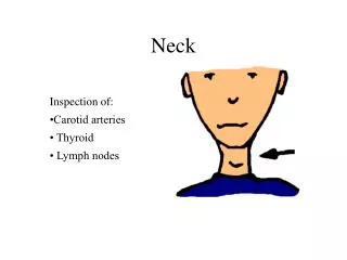
CLINICAL MANIFESTATIONS I-NECK PAIN C- Range of neck motion should include flexion, extension, rotation, and lateral bending. Normal flexion demonstrates the abillity to touch the chin to the chest. Normal neck extension allows the occiput to approach the prominent C7 spinous process. Rotation is normally 70 degrees bilaterally and lateral bending is 50 to 60 degrees bilaterally. Palpation for carotid artery pulses as well as for the presence or absence of supraclavicular adenopathy should be performed.
CLINICAL MANIFESTATIONS II- CERVICAL RADICULOPATHY A. Signs and symptom. Cervical radiculopathy implies pain traveling on the basis of an anatomic distribution to the shoulder or down the arm. Patients describe sharp pain and tingling or burning sensations in the involved area. There may be sensory or motor loss corresponding to the involved nerve root, and reflex activity may be diminished.
CLINICAL MANIFESTATIONS II- CERVICAL RADICULOPATHY B- Physical examination The shoulder abduction relief sign is characterized by having the patients place the palm ofhis hand flat onto the top of his skull; this causes symptomatic relief of the radicular pain Spurling’s test is performed by having the patient extnd the neck and rotate and laterlly bend the head toward the affected side; an axial compressiv forc is then applied to the top of the patient’ head. The test is positive when the maneuver reproduces the patients typical radicular arm pain.
CLINICAL MANIFESTATIONS II- CERVICAL RADICULOPATHY C- Herniation or degeneration of an intervertebral disc may produce spesific radicular patterns, depending on the level of involvement. Considerable overlap exist among the patterns outlined in the subsequent text. C5-6 and C6-7 are far more commonly involved tha C7-T1 or C4-5.
CLINICAL MANIFESTATIONS II- CERVICAL RADICULOPATHY C- Herniation or degeneration of an intervertebral disc 1. C5-6 (C6 nerve root affected). Pain will radiate to the shoulder or lateral arm and dorsal forearm. Anesthesia and paresthesias may be present in the thumb and index finger. Weakness, if present, will involve the biceps and wrist extensors. The brachioradialis or biceps reflex is often decreased or absent.
II- CERVICAL RADICULOPATHY C- Herniation or degeneration of an intervertebral disc 2. C6-7 (C7 nerve root affected). The pain distribution is similar to that of a C7 radiculopathy. Anesthesia and parestheias, when present, involve the ndex and log fingers. Weakness, if present, is noted in the ticeps, wrist flexors, and finger extensors. The triseps reflex may be reduced. CLINICAL MANIFESTATIONS
CLINICAL MANIFESTATIONS II- CERVICAL RADICULOPATHY C- Herniation or degeneration of an intervertebral disc 3. C7-T1 (C8 nerve root affected). Pain may occur along the medial aspect of the upper arm and forearm. Anesthesia and paresthesias involve the ring and small fingers. Weakness, if present, is notes in the finger flexors and intrinsic musculature of the hand. The triceps reflex may be reduced.
CLINICAL MANIFESTATIONS III- CERVICAL MYELOPATHY A. Signs and symptoms. Cervical myelopathy alone (e.g., in the absence of radiculopathy) is painless. This is due to the fact that there is spinal cord compression only. The pain becomes apparent only when compression of the spinal cord is accompanied by compression of the nerve root (myeloradiculopathy). Symptoms associated with spinal cord compression include gait disturbances with balance difficulty, fine motor dysfunction in the hands, and motor weakness. Bowel and bladder dysfunction is found late in the progression of cervical myelopathy. Physical findings often include difficulty with tandem gait, dysdiadochokinesia, hyperreflexia, and various sensory and motor changes.
CLINICAL MANIFESTATIONS III- CERVICAL MYELOPATHY B- Physical examination. Hoffmann’s reflex is often present, which is elicited by flicking the middle finger of the patient and observing forced finger and thumb interphalangeal joint flexion. There can be upgoing toes (e.g.,positive Babinski’s reflex) as well as associated clonus at the ankles. Myelopathy-related hand abnormalities include atrophy of the thenar musculature and an inability to maintain the ring and small fingers is an extended and adducted position (e.g., finger escape sign)Lhermitte’s sign involves flexion of the neck with an electric-shocklike sensation extending down the axial spine and/or extremities.In addition to the physical examination for neck pain, a thorough neurologic evaluation is necessary.This includes motor testing of all pertinent motor groups including the deltoid, biceps, tricepswrist flexors/extensors, finger flexors/extensors and the interossei.
CLINICAL MANIFESTATIONS III- CERVICAL MYELOPATHY B- Physical examination. Additionally, lower extremity strength needs to be tested including hip flexors, nee extensors and flexors, hip abductors and adductors, ankle dorsiflexors and plantar fleksors as well as the function of the function of the extensor hallucis longus, and peroneals. Sensory examination should include light touch, pinprick, and vibration sense using a tuning fork. Reflex examination should include the triceps, biceps and brachioradialis, quadriceps, and the Achilles tendon Another abnormal finding is the inverted radial reflex, characterized by spontaneous fnger flexion whenthe examiner attempts to elicit a brachioradialis reflex. Gait should be tested during normal gait as well as with toe to heel walking.
DIAGNOSTIC EVALUATION I- LABORATORY STUDIES. Laboratory studies should include routine blood workup; a complete blood count with differential, an erythrocyte sedimentation rate (ESR) and C-reactive protein(CRP). These results will most commonly be abnormal when an infectious or malignant process is involved.
DIAGNOSTIC EVALUATION II- ELRCTROPHYSIOLOGICAL TESTING An electrogram (EMG) may be helpful in defining a spesific anatomic level when nerve compression is present. Such a study may also be helpful in ruling out other neurlogic disorders including peripheral neuropathy. At times, a double-crush syndrome may exist when cervical radiculopathy can coexist with carpal tunnal syndrome.
IMAGING STUDIES I- PLAIN X-RAYS. A plain x-ray series should include an anterior/posterior view, a lateralview, and oblique views. Degeneration can often be noted within the disc spaces and the facet joints. There are often osteophytes noted along the area of the disc space, and foraminal narrowing can be noted on oblique views. Clinical correlation with patient symptoms is often poor in those older than 40. Instability has been define as greater than 3ç5 mm of translation or 11 degrees of angulation betweem adjacent vertebral segments.
IMAGING STUDIES II- MYELOGRAPHY. Myelography can be used to help evaluate nerve root compression as well as compression of the spinal cord. Root compression is manifested by an extradural filling defect with obliteration of the nerve root sleeve. Flattening of the spinal cord can be appreciated on the lateral view. In cases of severe compression, there will be complete obstruction of flow of the myelogram dye. In most clinical situations, this test has given way to the magnetic resonance imaging (MRI)
IMAGING STUDIES III- COMPUTED TOMOGRAPHY Computed tomography (CT) is helpful in evaluating the degee of foraminal stenosis caused by bony osteophytes. In combination with myelographyi it provides superior imaging compared to myelography alone. It permits the visualizaton the spesific levels (e.g, C6-7) and locaton (e.g., lateral recess and foraminal) of nerve root compression; filling defects allow or the determination of the extent of spinal cord compression. Measurement of the diameter of the spinal canal can be made to help define pre-existing stenosis. Individuals with an anteroposteior spinal canal diameter less than 13 mm are considered to have congenital cervical stenosis. In addition, patients with a cord-compression ratio (anteroposterior cord diameter divided by transverse cor diameter) less than 0.40 tend to have worse neurologic function.
IMAGING STUDIES IV- MRI MRI perhaps the primary imaging modality overall for cervical spine disorders. It provides excellent visualization of the spinal cord and soft tissues. Measurements of sagittal and axial canal diameters as well as cord-compression ratios can be calculated from an MRI.
DIFFERENTIAL DIAGNOSIS I- Differential diagnoses to consider with cervical disc disease are numerous. When a history of trauma is present, cervical sprain, traumatic injury to the brachial plexus, fracture, dislocation, or post-traumatic instability need to be considered. II- Inflammatory conditions including rheumatoid arthritis and ankylosing spondylitis can also present with cervical pathplogy. An infectious process including discitis, osteomyeltis, or soft tissue abscess (especially in light of a clinical history that includes fever or chills) must be ruled out.
DIFFERENTIAL DIAGNOSIS III- Tumors can be a cause of neck and upper extremity symptoms. These may include metastatic tumors, primary bone tumors, and tumors within the spinal cord. Additonally, tumors involving the upper lung (Pancoast’ tumor) may cause symptoms consistent with a C8 radiculopathy and/or a Horner’s syndrome. The presence of a history of weight loss, night pain, and present or past malignancy should increase the physician’s sensitivity to the possibility of a malignant tumor. IV- Shoulder disorders including rotator cuff disease, instability, and impingement may cause pain referred to the neck and can be confused with a C5 radiculopathy. More cmmonly, the neck refers pain to the soulder and may actually be associated with the development of frank shoulder pathology.
DIFFERENTIAL DIAGNOSIS V- Neurologic disorders such as the demyelinating disease, multipl sclerosis, as well as disease nvolving the anterior horn cells must be considered in the differential diagnosis. VI- Finally, many other conditions such as peripheral nerve entrapment syndromes, reflex sympathetic dystroph, thoracic outlet syndrome, as well as coronary artery disease with angina pectoris may stimulate radicular type symptoms. Pathology in the neck or shoulder may take those areas more likely sites to which visceral pain refers.
TREATMENT I- Conservative care isthe primary treatment of patients with neck pain, with or without radicular symptoms. Lifestyle modifications should be instituted to avoid activities that tend to create or aggravate neck and arm symptoms.
TREATMENT I- Conservative care A. Typical activities to modify include athletic activities, sitting at a desk with neck flexion (e.g.,reading and typing) for extended periods of time, and driving. An ergonomic assessment of the modern computerized office s often helpful in decreasing day-long stresses to the neck. A soft cervical collar can be used to limit motion and allow the spasm to settle down. The use of two or three pillows at night, in order to decrease reflux symptoms or breathing problems, exacerbate cervical spine problems and should be avoided. Therefore, use of a cervical pillow under the nape of the neck at night help decrease spasms and pain, as it tends to optimize the position of the neck during sleep. Other modalities such as moist hate and light massage may prove beneficial
TREATMENT I- Conservative care B- Use of medications including anti-inflammatory medications help decrease the amount of inflammation and prvide pain relief. In cases of severe pain, mild narcotics may be useful. Muscle relaxants may also help decrease the amount of spasm and allow or more comfortable periods of rest. Short courses of steroids are sometimes needed to control the inflammatory process.
TREATMENT I- Conservative care C- Physical therapy is often useful in the treatment of neck and radicular arm pain, once the phase of severe pain and radicular problems resolve. Modalities including traction, ultrasound, or diathermy can give pain relief. Once the patient’s symptoms have begun to decrease, an exercise regimen can be added taking note that this does not exacerbate the neck or arm pain symptomsç Active ROM exercises along with some isometric exercises can help regain the strength of the neck.
TREATMENT II- Surgery is indicated in cases of significant radicular pain that has failed to respond to conservative treatment, or in the presence of significant neurologic deficits. Only a small percentage of patients with cervical spine problems eventually require surgery. However, if considered necessaryi the surgical procedure is either an anterior cervical discectomy and fusion or a posterior laminoforaminotomy. For cases of melopathy with significant disability, surgery can be a reasonable alternative. The goal of surgery with myelopathy is to prevent progression of the disease.
TREATMENT II- Surgery Postoperatively, some patients show improveent from their preoperative neurologic status. For myelopathy, surgery cnsists of either multipl anterior cervical discectomies/corporectomies and fusion versus posterior procedures such aslaminectomy alone, lamioplasty, or laminectomy and fusion. A small percentage of patients with significant multilevel disease or poor bone quality are good candidates for a combined anterior/posterior procedureç Surgery should be done emergently in the setting of anepidural abscess.
PROGNOSIS I- The prognosis for patients with axial neck pain is, in general, good. In a folow-u of a seies of 205 patients with neck pain and treated nonoperatively, 79% were noted to be either asymptomatic or improved at 10-year follow-up,13% were noted to be unchanged, and 8% were felt to have worsening symptoms. Surgery for axial neck pain by itself is rarely indicated, except perhaps in the setting of instability.
PROGNOSIS II- The prognosis for patients with cervical radiculopathy is also, in general, favorable. A significant number of patients tendto respond to nonoperative measures and show significant improvement 2 to 3 months after the onset of symptoms. A series of 26 patients with cervical disc herniation and radiculopathy were managed nonoperatively with traction, medications and education. A1 year follow-up show succesful nonoperative management 24 of the26 patients. For patients who have radicular symptoms despite 2 to 3 months of nonsurgical treatment, or who have significant weakness, surgery is a reasonable option. The prognosis for improvement with surgery is generally favorable. Most patients experience significant improvement in their radicular painç
PROGNOSIS III- Cervical myelopathy with early myelopathy and no significant neurologic deficits can initially be followed in an outpatient setting. The prognosis for cervical myelopathy in general shows that a high percentage of these patients slowly deteriorate over time. The deteioration is often slow and occurs over years; a small percent of cases may display signs and symptoms of rapid progression. In patients with gross findings of myelopathy with significant cord compression and impairment, surgery is a reasonable option. The goal of surgery is to prevent deterioration and potentially promote improvement in their overall neurologic status. In a series of patients treated surgically for cervical myelopathy, 90% of patients had significant neurologic improvement and 80% has significant pain relief.