Download
1 / 21
280 likes | 878 Views
Neck Swellings. Causes of a lump in the Neck. Congenital Cysts. Lymph Node Enlargement. Salivary Gland Disorders. Lumps in the Skin. Thyroid Diseases. Parathyroid Tumour. Thymus Tumour. Lymph Node Disorders 1.

E N D
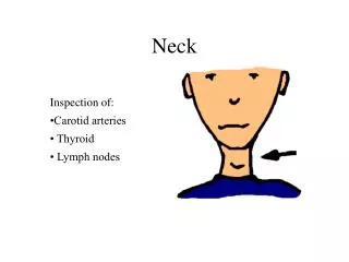
Causes of a lump in the Neck • Congenital Cysts. • Lymph Node Enlargement. • Salivary Gland Disorders. • Lumps in the Skin. • Thyroid Diseases. • Parathyroid Tumour. • Thymus Tumour.
Lymph Node Disorders 1 • Patients are often referred to surgeons for biopsy of a lymph node in the cervical region. • Nodes become palpable when their diameter exceeds 1 cm, but impalpable nodes may contain tumour! • Often there are no other symptoms or signs. • Tender nodes are usually inflammatory, whereas non-tender nodes may be malignant. • Isolated lymph node enlargement may be caused by local disease within its field of drainage. Alternatively, an enlarged cervical may be a part of systemic lymphoadenopathy.
Lymph Node Disorders 2 • Infective • Bacterial (pyogenic infections in drainage area)→ e.g. TB • Viral → e.g. HIV • Protozoal→ Toxoplasmosis • Neoplasm • Lymphoma • Metastastic squamous carcinoma • Other meastastic tumours • Secondary (Metastatic Tumour) • Squamous carcinoma or melanoma of the skin of the neck
Lymph Node Disorders 3 • Cervical TB: • The causative organism is human or bovine (e.g. drinking milk from cattle infected with TB). • Occasionally the disease is secondary to an active pulmonary infection. • Clinical Features : Small discrete nodes → Enlarge, become matted together & caseate → Formed Abcess eventually burst throught the deep fascia into the subcutaneous tissues. • X-ray of the neck: Chronic TB nodes show flecks of calcification.
Lymph Node Disorders 4 • Lymphoma: • An enlarged cervical lymph node is a common presentation of non-Hodgkin’s lymphoma or Hodgkin’s disease. • The disease may be at an early stage and there may be no other symptoms or clinical signs. • The diagnosis is then made by histological examination of a biopsy speciment.
Lymph Node Disorders 5 • Metastatic Cancer • When cervical adenopathy is noted, the upper aerodigestive tract must be carefully examined to exclude a tumour. • This usually involves direct examination of the mouth, pharynx, larynx and esophagus. • When inspection of these areas does not reveal an abnormality, palpation of the tonsils and tongue may reveal an occult tumour. • Random biopsies from the nasopharynx may also reveal an unsuspected tumour.
Lymph Node Disorders 6 • The head and neck, and in particular the oral cavity, nose, pharynx and larynx, must be examined to detect a primary cause of lymphadenopathy. • Fine-needle aspiration cytology can be diagnostic for secondaryhead and neck tumors or lymphoma. • CT scanning helps to find the extent of the lymphadenopathy. • Painless neck nodes in patients over 45 are often due to metastasis from carcinoma. In modt cases, the primary is within the head and neck. Such nodes must be biopsied in the first instance; a thorough search for a primary lesion is the key to diagnosis.
Salivary Glands Disorders 1 • Salivary Gland Tumours • Enlarging mass anterior/inferior to ear or at the mandible angle is suspect • Benign • Asymptomatic except for mass • Malignant • Rapid growth, skin fixation, cranial nerve palsies
Salivary Glands Disorders 2 • Salivary Gland Stone Disease (Sialolithiasis) • Calculi cause salivary gland swelling by blocking the duct. • Rarely cause complete obstruction, but the patient usually experiences intermittent swelling or pain at meal times when the salivary flow is high. • Some but not all stones are radio-opaque →X-ray for diagnosis? • Calculi may pass spontaneously, but are more likely to remain in situ.
Lumps in the Skin • Sebaceous cysts • Characterized by the presence of a punctum • Furuncles or boils • Infection in hair follicles • If the infection spreads to involve the dermis and subcutaneous tissue → carbuncle. • Kaposi’s sarcoma • In patients with AIDS.
Thyroid Diseases 1 • Inflammatory • De Quervain’s thyroiditis • Riedel’s Thyroiditis • Autoimmune • Hashimoto’s thyroiditis • Classification of Goitres: • Simple (non-toxic Goitre) • Simple Hyperplastic Goitre • Multinodular Goitre • Toxic Goitre • Diffuse Goitre (Grave’s disease) • Toxic Nodule • Toxic Multinodular Goitre • Neoplastic Goitre • Benign • Adenoma • Malignant • Papillary • Follicular • Anaplastic • Medullary
Thyroid Diseases 2 • Simple Goitre: • Simple Hyperplastic: • Pathologically → Iodine Deficiency. Physiologically → Puberty & Pregnancy. • T4 is N. TSH is ↑ • CXR and thoracic inlet radiograph to exclude tracheal compression or deviation. • CT scan • Multinodular: • Dysnea & Dysphagia if the gland is very large. • Pain may occur due to hemorrhage into a cyst. • Free T4, TSH. • An isotope scan will identify toxic nodules. • Radiograph thoracic inlet.
Thyroid Diseases 3 • Toxic Goitre • TSH receptor antibodies are present. • Nodules function independently of TSH stimulation. • ↓TSH, ↑T3, ↑Free T4 • Isotope Scan may show hot nodules. • Neoplastic Goitre(pts. Are usually euthyroid- diagnosed by US - FNA - CXR) • Benign: • Adenomas: • True follicular adenomas are encapsulated and usually greater that 2 cm on presentation. • Solid on ultrasound and ‘cold’ on isotope scans. • FNA will not confirm their nature. They must be excised in total and examined by the pathologist before confirming their benign nature. • Malignant • Well Differentiated: • Papillary (60%) • Follicular (20%) • Anaplastic Carcinoma (5-8%) • Medullary (5%)
Parathyroid Tumours • Usually Single. • The lower glands are affect more commonly than the upper glands. • Most are benign adenomas and carcinoma of the parathyroid is rare. • May coexist with other endocrine tumors. Such as:………… &……………..In……… & ……………&……………. In …………... • ↔ Hyperparathyrodism
Thymus Disorders • Thymus Gland Controls the development of T lymphocytes in the Embryo and Neonate. • In adults it is a fat-infiltrated remnant. • Rare site of mediastinal Tumour which can arise from either: • The epithelium • Lymphoid tissue • Both • They can be: • Benign (occasionally cystic) • Malignant (and rapidly invasive) • Peak incidence in the sixth and seventh decades. • Clinical Features: • A mediastinal mass. • Associated with Myasthenia Gravis. • Associated with an immune deficiency state.
Thanks For Your Patience Wish you a Happy Weekend