Download

1 / 22
220 likes | 225 Views
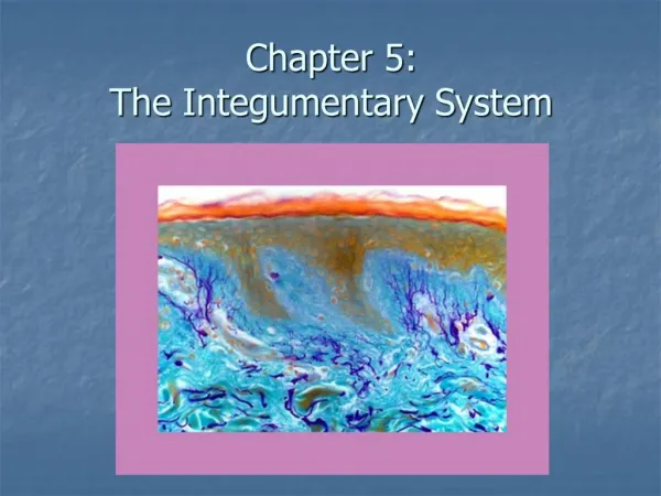
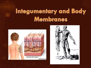
Chapter 5 The Integumentary System and Body Membranes. CLASSIFICATION OF BODY MEMBRANES. Classification of body membranes (Figure 5-1) Epithelial membranes—composed of epithelial tissue and an underlying layer of connective tissue

E N D
CLASSIFICATION OF BODY MEMBRANES • Classification of body membranes (Figure 5-1) • Epithelial membranes—composed of epithelial tissue and an underlying layer of connective tissue • Connective tissue membranes—composed largely of various types of connective tissue
CLASSIFICATION OF BODY MEMBRANES • Epithelial membranes • Cutaneous membrane—the skin • Serous membranes—simple squamous epithelium on a connective tissue basement membrane • Types • Parietal—line walls of body cavities • Visceral—cover organs found in body cavities • Examples • Pleura—parietal and visceral layers line walls of thoracic cavity and cover the lungs • Peritoneum—parietal and visceral layers line walls of abdominal cavity and cover the organs in that cavity • Diseases • Pleurisy—inflammation of the serous membranes that line the chest cavity and cover the lungs • Peritonitis—inflammation of the serous membranes in the abdominal cavity that line the walls and cover the abdominal organs
CLASSIFICATION OF BODY MEMBRANES • Epithelial membranes (cont.) • Mucous membranes • Line body surfaces that open directly to the exterior • Produce mucus, a thick secretion that keeps the membranes soft and moist
CLASSIFICATION OF BODY MEMBRANES • Connective tissue membranes • Do not contain epithelial components • Produce a lubricant called synovial fluid • Examples are the synovial membranes in the spaces between joints and in the lining of bursal sacs
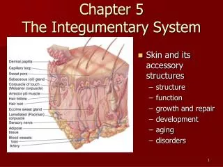
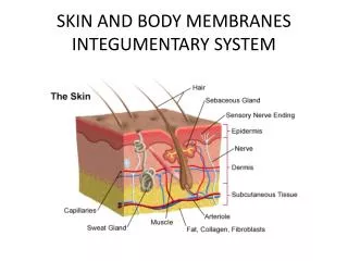
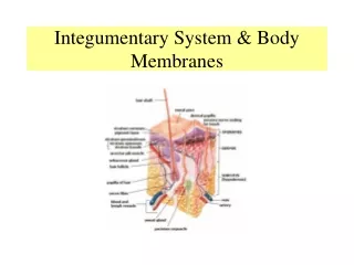
THE SKIN • Structure (Figure 5-2)—two primary layers called epidermis and dermis • Epidermis • Outermost and thinnest primary layer of skin • Composed of several layers of stratified squamous epithelium • Stratum germinativum—innermost layer of cells that continually reproduce; new cells move toward the surface • As cells approach the surface, they are filled with a tough, waterproof protein called keratin; eventually cells flake off of body • Stratum corneum—outermost layer of keratin-filled cells • Skin pigment—produced by deepest epidermal layer; gives color to the skin • The brown pigment melanin is produced by specialized cells in deepest epidermal layer • Blisters—caused by breakdown of union between cells or primary layers of skin
THE SKIN • Structure (cont.) • Dermal-epidermal junction—specialized area between two primary skin layers • Dermis • Deeper and thicker of the two primary skin layers; composed largely of connective tissue • Upper papillary layer of dermis characterized by parallel rows of tiny bumps called dermal papillae • Ridges and grooves in dermis form pattern unique to each individual • Basis of fingerprinting • Improves grip for tool use and walking • Deeper reticular layer of dermis filled with network of tough, interlacing, collagenous and stretchable elastic fibers • Number of elastic fibers decreases with age and contributes to wrinkle formation • Dermis also contains nerve endings, muscle fibers, hair follicles, sweat and sebaceous glands, and many blood vessels
THE SKIN • Accessory structures of the skin • Hair (Figures 5-5) • Lanugo—soft hair of fetus and newborn • Hair follicle—epidermal tubelike structure required for hair growth • Hair papilla—structure from which hair growth begins • Hair root—lies hidden in follicle • Hair shaft—visible part of hair • Arrector pili—specialized smooth muscle that produces “goose bumps” and causes hair to stand up straight
THE SKIN • Accessory structures of the skin (cont.) • Receptors (Figure 5-2) • Specialized nerve endings—make it possible for skin to act as a sense organ • Meissner’s corpuscle—capable of detecting light touch • Pacinian corpuscle—capable of detecting pressure
THE SKIN • Accessory structures of the skin (cont.) • Nails (Figure 5-6) • Produced by epidermal cells over terminal ends of fingers and toes • Nail body—visible part of nail • Root—lies in a groove; hidden by cuticle • Lunula—crescent-shaped area nearest root • Nail bed may change color with change in blood flow
THE SKIN • Accessory structures of the skin (cont.) • Skin glands—two types • Sweat, or sudoriferous, glands • Eccrine sweat glands • Most numerous, important, and wide-spread of the sweat glands • Produce perspiration or sweat, which flows out through pores on skin surface • Function throughout life and assist in body heat regulation • Apocrine sweat glands • Found primarily in axilla and around genitalia • Secrete a thicker secretion quite different from eccrine perspiration • Breakdown of secretion by skin bacteria produces odor • Sebaceous glands • Secrete oil or sebum for hair and skin • Level of secretion increases during adolescence • Amount of secretion is regulated by sex hormones • Sebum in sebaceous gland ducts may darken to form a blackhead
THE SKIN • Skin cancer • Functions of the skin • Protection—first line of defense against: • Infection by microbes • Ultraviolet rays from sun • Harmful chemicals • Cuts and tears
THE SKIN • Functions of the skin (cont.) • Temperature regulation • Skin can release almost 3000 calories of body heat per day • Mechanisms of temperature regulation • Regulation of sweat secretion • Regulation of blood flow close to the body surface • Sense organ activity • Skin functions as an enormous sense organ • Receptors serve as receivers for the body, keeping it informed of changes in its environment
THE SKIN • Burns • Treatment and recovery or survival depend on total area involved and severity or depth of the burn • Body surface area is estimated using the “rule of nines” (Figure 5-8) in adults • Body is divided into 11 areas of 9% each • Additional 1% located around genitals
THE SKIN • Burns (cont.) • Classification of burns • First-degree (partial-thickness) burns—only the surface layers of epidermis involved • Second-degree (partial-thickness) burns—involve the deep epidermal layers and always cause injury to the upper layers of the dermis • Third-degree (full-thickness) burns—characterized by complete destruction of the epidermis and dermis • May involve underlying muscle and bone • Lesion is insensitive to pain because of destruction of nerve endings immediately after injury—intense pain is soon experienced • Risk of infection is increased