Download
1 / 28
280 likes | 289 Views
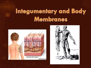
Body Membranes and Integumentary System. Body Membranes: Thin, sheet-like organs that cover surfaces and line cavities 4 Types Serous: line body cavities that lack any opening to outside of body Simple squamous epithelium and thin layer of loose connective tissue under Secretes serous fluid

E N D
Body Membranes: Thin, sheet-like organs that cover surfaces and line cavities • 4 Types • Serous: line body cavities that lack any opening to outside of body • Simple squamous epithelium and thin layer of loose connective tissue under • Secretes serous fluid • Pericardial, pleural, peritoneum • Mucous: line body cavities and tubes that open to outside • EX: lining of urinary and digestive system, respiratory and reproductive systems
Synovial: form the inner linings of freely moveable (synovial) joins • Comprised of a layer of loose connective tissue with adipose • Only body membrane with no epithelium • Secrete thick, colorless fluid (synovial) for lubrication and transport • Cutaneous: skin plus accessory organs is called the Integumentary system
Integumentary System • Function of the Skin • Provides protective covering • Retards water loss • Regulates body temperature • Has various sensory receptors (pain, pressure, etc) • Synthesizes chemicals • Secretion of some waste products (sweat)
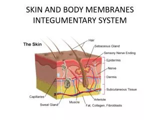
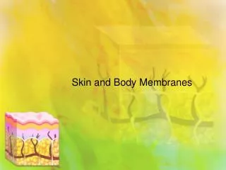
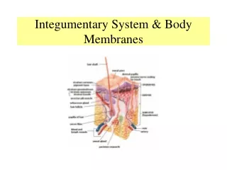
Three Layers of Skin • Epidermis: outer, stratified squamous epithelium • Dermis: Deeper, thicker than epidermis • Comprised of fibrous connective tissue, smooth muscle tissue, nerve fibers, blood vessels, infolded epithelium • Separated by basement membrane • Hypodermis: Layer under dermis • Also called subcutaneous layer • Composed of loose connective tissue with abundance of adipose • Functions to bind skin to underlying structures
Epidermis • Divided into 5 layers or strata • Deepest to most external • Stratum Basale (basal layer) • Stratum Spinosum (spiny layer) • Stratum Granulosum (granular layer) • Stratum Lucidum (clear layer) • Statum Corneum (horned layer) (dandruff)
Epidermis Deepest layer is called Stratum Basale or stratum germinatium. These cells are actively dividing • The older cells are pushed outward towards surface, away from their nutrients • Gradually flatten • Keratinize, dry out, harden and die • Outermost layer of dead keratinized cells is called stratum corneum
Epidermis Cont. • Thickness of Stratum Corneum varies • Thickness in soles of feet, and palms of hands • Division rate increases where rubbing or pressing on the skin occurs • Callouses on hand, corns on feet • These cells are called keratinocytes • Melanocytes: scattered in the stratum basale and produce melanin. This is a dark pigment • Absorbs visible and ultraviolet light • Purpose is to protect deeper cells from UV light
Dermis • Gives skin toughness and elasticity • Muscle fibers within • Smooth: associated with hair follicles (arrector pili) and glands • Skeletal: anchored in the dermis of the face for facial expressions • Blood Vessels: nutrients to dermis and epidermis • Nerve Fibers: • Motor: neurons that convey from CNS to effectors at the body (muscles and glands) • Sensory: convey from periphery of body to CNS • Ex: temp. and tissue damage receptors (pain) • Pressure receptors: • - pacinian corpuscles: deep and stimulated by heavy pressure • - meissners corpuscles: in dermal papillae and stimulated by light pressure or touch
Dermis Cont. • Epithelial Derived Excessory Organs • Hair follicles • Sebacous glands • Sweat glands
Subcutaneous (Hypodermis) • Loose connective tissue with high amount of adipose • Blood vessels to dermis • Purpose is to bind skin to underlying organs • Adipose within also functions as a heat insulator and in cushioning the body
Skin Cancers • Cutaneous Carcinoma • Originates from deeper epithelial cells • Most common skin cancer • Risk increased by prolonged exposure to moderate levels of UV light • Slow growing, doesn’t deepen rapidly, often curable
Skin Cancers • Cutaneous Melanoma • Develops from melanocytes • Very darkly pigmented • Risk increased by intermittent exposure to intense levels of UV light (tanning beds!!!) • Thicken and metastasizes quickly • More deadly than cutaneous carcinoma
Accessory organs in skin • Hair Follicles: on all skin surfaces except palms, soles of feet, parts of reproductive organs • Shaft: part above the surface • Root: part beneath surface • Expanded base called bulb • Bulb has medial indention called papilla • Well nourished cells around papilla divide rapidly and push up older cells above them that then flatten, keratinize, increase number of desosomes, dryout and die
Baldness: quit replacing hairs, is a sex linked trait • Color: largely determined by pigments produced by melanocytes • Arrector Pili muscle: smooth muscles attached at one end near bottom of root and run oblique to attach near basement membrane at other end • If frightened or cold- contract elevating hair “goose bumps”
Sebaceous glands: “oil glands” empty into hair follicles • Holocrine secretors: secrete sebum (oily) • Purpose is to keep skin and hair soft and pliable and waterproof • Responsible for acne- when ducts become inflamed and blocked • Nails • Function as protection • Composed of • Nail plate: dead keratinized cells fused • Nail bed: special epithelium (divides rapidly) • Lunula: white ½ moon shape in bed at base. Most active cell division
Sweat (sudoriferious) glands • Coiled tubular gland • Secretes serous solution (sweat) • Sweat is water, electrolytes, and waste products • 2 types of sweat glands • Apocrine: apocrine secretion, terminal end is dumped in lumen • Has fair amount of odor • Scent glands in animals • Stimulated by emotions • Associated with hair follicles • Found in axilla and groin
Eccrine: Secrete by merocrine exocytosis • not associated with hair follicles • Stimulated by increase in body temp.
Body Temp. Regulation • Temperature (heat) affects the reaction rates n body • Normally 98.6°F/37°C • Most endogenous body heat is produced as a product of cellular respiration • Most significant tissue is skeletal muscle
Loss of body heat to environment • Radiative (radiation) • Majority of heat loss by this • Conduction • One molecule bumping into another • Convection • Hot air is lighter than cold air • Evaporation • Change from liquid to gas
CLINICAL APPLICATION • Heat Exhaustion: due to electrolyte imbalance within body fluids due to excess sweat • Dizziness, headache, muscle cramps, nausea • Treat with water and electrolytes
Skin Color • Genetic Factors • Genes regulate amount of melanin produced per melanocyte • Albinism: genetic mutation inherited in one or more of the genes leading to melanin production • NO MELANIN • Environmental Factors • Ultra violet light stimulates melanocytes to increase melanin production and cytocrine transfer
Skin color cont. • Physiological factors • Increase in dermal blood flow: pinkish • Oxygenated hemoglobin: bright red • Deoxygenated Hemoglobin: dull, brick red • Decreased dermal blood flow: bluish (cyanotic) because of increase in deoxygenated hemoglobin (when cold) • Diseases: • Pulmonary diseases • Jaundice: yellowish coloration due to high amount of bile pigments in body fluids