Download

1 / 53
640 likes | 1.21k Views
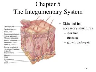
Chapter 5 The Integumentary System. Introduction. The organs of the integumentary system include the skin and its accessory structures including hair, nails, and glands, as well as blood vessels, muscles and nerves

E N D
Introduction • The organs of the integumentary system include the skin and its accessory structures including hair, nails, and glands, as well as blood vessels, muscles and nerves • Dermatology is the medical specialty for the diagnosis and treatment of disorders of the integumentary system.
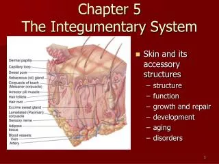
Structure of the Skin • The skin (cutaneous membrane) covers the body and is the largest organ of the body by surface area and weight • Its area is about 2 square meters (22 square feet) and weighs 4.5-5kg (10-11 lb), about 7% of body weight • It is 0.5 – 4 mm thick, thinnest on the eyelids, thickest on the heels; the average thickness is 1 – 2 mm
Structure of the Skin • It consists of two major layers: • outer, thinner layer called the epidermis, consists of epithelial tissue (see video) • inner, thicker layer called the dermis • Beneath the dermis is a subcutaneous (subQ) layer (also called hypodermis) which attaches the skin to the underlying tissues and organs.
Structure of the Skin • The epidermis has a number of important characteristics: • the epidermis is composed of keratinized stratified squamous epithelium • it contains four major types of cells: • Keratinocytes (90% of the cells) produce keratin which is a tough fibrous protein that provides protection
Structure of the Skin • Melanocytes: which produce the pigment melanin that protects against damage by ultraviolet radiation • Langerhans cells: involved in immune responses, arise from red bone marrow • Merkel cells: which function in the sensation of touch along with the adjacent tactile discs
Epidermis • The epidermis contains four major layers (thin skin) or five major layers (thick skin) • Stratum basale (deepest layer) or stratum germinativum, where continuous cell division occurs which produces all the other layers • Stratum spinosum, 8-10 layers of keratinocytes • Stratum granulosum, which includes keratohyalin and lamellar granules
Epidermis • Stratum lucidum is present only in thick skin (the skin of the fingertips, palms, and soles) • Stratum corneum: composed of many sublayers of flat, dead keratinocytes called corneocytes or squames that are continuously shed and replaced by cells from deeper strata; constant friction can stimulate formation of a callus. • Keratinization, the accumulation of more and more protective keratin, occurs as cells move from the deepest layer to the surface layer • Dandruff - an excess of keratinized cells shed from the scalp
Dermis • The dermis has several important characteristics: • is composed of connective tissue containing collagen and elastic fibers • contains two layers • the outer papillary region consists of areolar connective tissue containing thin collagen and elastic fibers, dermal papillae (including capillary loops),corpuscles of touch and free nerve endings
Dermis • The deeper reticular region consists of dense irregular connective tissue containing collagen and elastic fibers adipose cells, hair follicles, nerves, sebaceous (oil) glands, and sudoriferous (sweat) glands • Striae or stretch marks can appear if the skin is stretched too much
Dermis • Lines of cleavage - “tension lines” in the skin indicate the predominant direction of underlying collagen fibers • Epidermal ridges reflect contours of the underlying dermal papillae and form the basis for fingerprints (and footprints); their function is to increase firmness of grip by increasing friction. • Dermatoglyphics - the study of the pattern of epidermal ridges
Cleavage (Tension) Lines and Striae • Cleavage (tension) lines: elastin and collagen fibers oriented in some directions more than in others • Important in surgery • If incision parallel to lines, there is less gapping, faster healing, less scar tissue • If skin is overstretched, striae (stretch marks) occur
Structural Basis of Skin Color • Variations in skin color arise from variations in the amounts of three pigments: melanin, carotene, and hemoglobin • Melanin - a yellow-red or brown-black pigment produced by melanocytes (located mostly in the epidermis, where it absorbs UV radiation) • The amount of melanin causes the skin’s color to vary from pale yellow to red to tan to black • The number of melanocytes are about the same in all people; differences in skin color is due to the amount of pigment produced
Structural Basis of Skin Color • A benign localized overgrowth of melanocytes is a nevus or mole • Albinism is an inherited inability to produce melanin - vitiligo is a condition in which there is a partial or complete loss of melanocytes from patches of skin • Carotene - yellow-orange pigment(found in the stratum corneum, dermis, and subcutaneous layer) • Hemoglobin - red color (located in erythrocytes flowing through dermal capillaries)
Subcutaneous Layer • Subcutaneous (subQ) layer (also called hypodermis) is not part of the skin but, among its functions, it attaches the skin to the underlying tissues and organs; this layer (and sometimes the dermis) contains lamellated (pacinian) corpuscles which detect external pressure applied to the skin.
Accessory Structures of the Skin • include hair, skin glands, and nails • Hairs (pili) have a number of important functions: • protection • reduction of heat loss • sensing light touch
Accessory Structures of the Skin - Hair • Hair is composed of dead, keratinized epidermal cells • Hair consists of: • shaft which mostly projects above the surface of the skin • root which penetrates into the dermis • hair follicle • epithelial root sheath – (downward continuation of the epidermis) • dermal root sheath -
Accessory Structures of the Skin • There are different types of hairs including lanugo, vellus hairs and terminal hairs • Hair color is determined by the amount and type of melanin • Sebaceous (oil) glands are connected to hair follicles
Skin Glands • Sebaceous glands secrete an oily substance called sebum which prevents dehydration of hair and skin, and inhibits growth of certain bacteria (Sebum=triglycerides, cholesterol, proteins, and inorganic salts) • Sudoriferous (sweat) glands-- 2 types: • Eccrine sweat glands • Apocrine sweat glands
Sudoriferous (Sweat) Glands • Numerous eccrine (or merocrine) sweat glands helps to cool the body by evaporating, and also eliminates small amounts of wastes • Apocrine sweat glands, located mainly in the skin of the axilla, groin, areolae, and bearded facial regions of adult males. • their excretory ducts open into hair follicles- this sweat is secreted during emotional stress and sexual excitement.
Ceruminous Glands • Modified sweat glands located in the ear canal • Along with nearby sebaceous glands, they are involved in producing a waxy secretion called cerumen (earwax) which provides a sticky barrier that prevents entry of foreign bodies into the ear canal.
Nails • Nails are composed of hard, keratinized epidermal cells located over the dorsal surfaces of the ends of fingers and toes • Each nail consists of: • free edge • transparent nail body (plate) with a whitish lunula at its base • nail root embedded in a fold of skin
Types of Skin • There are two major types of skin: • thin (hairy) skin covers all body regions except the palms, palmar surfaces of digits, and soles • thick (hairless) skin covers the palms, palmar surfaces of digits, and soles
Deep Wound Healing • Injury extends into dermis & hypodermis • Scar tissue is formed • Some normal function lost • Occurs in four phases • Inflammatory phase • Migratory phase • Proliferative phase • Maturation phase
Inflammatory Phase • Blood clot forms loosely uniting wound edges • Inflammation occurs • Eliminates microbes, foreign material, and dying tissue • Increases diameter of local blood vessles • Enhancing delivery of nutrients, immune cells, and fibroblasts
Migratory Phase • Clot dries into scab • Epithelial cells migrate beneath scab and bridge wound • Fibroblasts migrate and lay down collagen fibers and glycoproteins in dermis • New blood vessels grow • Tissue called granulation tissue during this phase destined to become scar tissue
Proliferative Phase • Extensive growth of epithelium • Deposition of collagen in random patterns by fibroblasts • Continued growth of blood vessels
Maturation Phase • Scab sloughs off once epidermis restored to normal thickness • Granulation tissue developing into scar tissue • Fibroblasts decrease in number • Blood vessels restored to normal • Scar tissue formation called fibrosis • Elevated scars called • Hypertrophic scars • If contained within sight of original wound • Keloid scars • If extended beyond original wound
Aging and the Integumentary System Effects: • wrinkling • decrease of skin’s immune responsiveness • dehydration and cracking of the skin • decreased sweat production • decreased numbers of functional melanocytes resulting in gray hair and atypical skin pigmentation • loss of subcutaneous fat • a general decrease in skin thickness • an increased susceptibility to pathological conditions • Growth of hair and nails decreases; nails may also become more brittle with age.
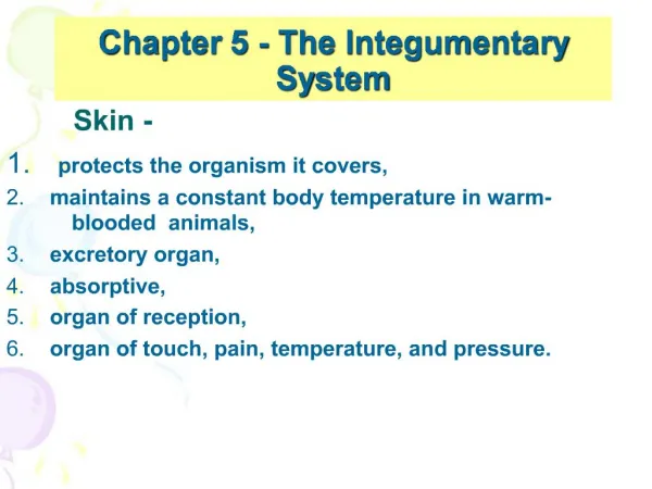
Functions of the Skin • regulation of body temperature • blood reservoir • protection • cutaneous sensations • excretion and absorption • synthesis of vitamin D