Download
1 / 44
440 likes | 454 Views
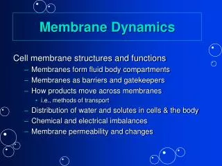
Membrane Dynamics cont’d. How many substrates can a carrier move? Active Transport Secondary Active Transport Transepithelial transport. How many substrates can a carrier move?. How many substrates can a carrier move?. Direction of substrate movement.

E N D
Membrane Dynamics cont’d • How many substrates can a carrier move? • Active Transport • Secondary Active Transport • Transepithelial transport
Carrier mediated transport into cells:- net movement as long as there is a concentration gradient across the mb Facilitated Diffusion Diffusion and carrier proteins
Function during disequilibrium Ca++ Ca++ Ca++ Calcium entry
Some molecules need to be in disequilibrium. • Levels of extracellular calcium and extracellular sodium (Na+) are high. Na+ Ca++ Na+ Ca++
Active Transport • Na+ is removed from cells against its concentration gradient • Need ATP energy for this work
Active Transport: Na+/K+ ATPase Na+ K+ Low levels of intracellular Na+
Active Transport: Na+/K+ ATPase 1. 3 Intracellular Na+ Ions bind Onto Na+/K+ ATPase
Active Transport: Na+/K+ ATPase 2. ATP hydrolysis
Active Transport: Na+/K+ ATPase • 3. The 3 Na+ ions are released Into the ECF
Active Transport: Na+/K+ ATPase • 4. Binding of 2 K+ ions from ECF
Active Transport: Na+/K+ ATPase • 5. Intracellular release of 2K+ ions
Secondary (indirect) Active Transport Symport driven by Na+ concentration gradient for trans-epithelial transport,
Transepithelial transport: • Primary active transport • Secondary active transport • Facilitated diffusion Must have low levels of intracellular Na+ To drive transepithelial transport
Intracellular glucose provide energy for primary and secondary active transport.
Where does transepithelial transport occur? • Glucose absorption in the intestine • Glucose absorption in the nephron • Glucose is moved from the mucosal surface of the epithelium to the serosal surface. • Glucose is moved from the apical surface of the cells to the basal surface of the cells.
How does water move in the body? • The cell membrane is semi-permeable • Water can move freely • Water is in equilibrium between cells and extracellular fluids (osmotic equilibrium) • Ions and solutes are disequilibrium • Osmosis water moves along its concentration gradient across a semi-permeable membrane
Distribution of solutes in the body fluid compartments plasma Interstitial fluid Intracellular fluid
Distribution of solutes in the body fluid compartments Ions and solutes are in disequilibrium
Ions and solutes are in disequilibrium • Water can cross the cell membrane Na+ K+ Na+ K+ proteins
Osmosis • water moves along its concentration gradient across a semi-permeable membrane • Water moves to dilute a solute
Osmotic pressure is pressure exerted to counter the movement of water to dilute something
Osmolarity • Describes the number of particles in solution • Know this and the direction of water movement can be predicted • # of particles in 1 liter of solution • Is expressed as osmoles/L, or OsM • If very dilute: milliosmoles/L, or mOsM • Human body, approx 300 mOsM
Osmolarity: number of particles in 1L • 1 M glucose = 1 OsM glucose • 1M NaCl = 2 OsM NaCl, because NaCl disassociates to 2 ions in solution. Na+ Cl-
Solution A 1 OsM glucose A is hyposmotic to B (A has fewer particles than B) Solution B 2 OsM glucose B is hyperosmotic to A (B has more particles than A) Compare the osmolarity of 2 solutions:
Solution C 1 OsM NaCl C is hypotonic to B (C has fewer particles/L than B) Compare the osmolarity of 2 solutions: • Solution B • 2 OsM glucose • B is hyperosmotic to C • (B has more particles/L than A)
Compare the osmolarity of 2 solutions: • Solution A • 1 OsM glucose • A is isosmotic to C • Solution C • 1 OsM NaCl • C is isosmotic to A
Osmosis, the diffusion of water across the cell membrane, has consequences on cells • After water leaves a cell, the volume changes (it can shrink)
Tonicity • Describes how the cell volume will change in a solution
P is penetrating solute N is nonpenetrating solute
Cell gains volume in a hypotonic solution • Cell looses volume in a hypertonic solution • Cell keeps the same volume in an isotonic solution.
Tonicity indicates how the cell volume will change in a solution • In a hypotonic solution, the cell has a higher concentration of a nonpenetrating solute than the solution, water moves in. • In a hypertonic solution, the cell has a lower concentration of nonpenetrating solute than the solution, water leaves the cell
During intavenous injection: • 0.9% (normal) saline isotonic • D5--.9% saline (5% dextrose) isotonic • D5W hypotonic • 0.45% saline hypotonic • Vs dehydration hypotonic • Vs blood loss isotonic