Download
1 / 58
1.06k likes | 2.45k Views
Introduction to Virology. Learning Objectives: On completing this session, you should be able to: Understand what a virus is & explain how viruses differ from all other organisms. Summarize the history of virology & explain how the present state of our knowledge of viruses was achieved.

E N D
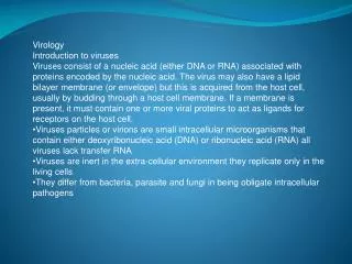
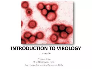
Introduction to Virology Learning Objectives: On completing this session, you should be able to: • Understand what a virus is & explain how viruses differ from all other organisms. • Summarize the history of virology & explain how the present state of our knowledge of viruses was achieved. • Describe the techniques most frequently used to study viruses. Joko Pamungkas
Part 1 Joko Pamungkas
Virus diversity • More biological diversity within viruses than in all the rest of the bacterial, plant & animal kingdoms put together. • This results from the success of viruses in parasitizing all known groups of living organisms. • Understanding this diversity is the key to comprehending the interactions of viruses with their hosts. Joko Pamungkas
Viruses are distinct from living organisms • Viruses are submicroscopic, obligate intracellular parasites. • However, a few groups of prokaryotic organisms that have specialized intracellular parasitic life-cycles & which confound the above definition - the Rickettsiae & Chlamydiae - obligate intracellular parasitic bacteria which have evolved so that they can exist outside the cells of their hosts only for a short period of time before losing viability. • Therefore it is necessary to add further clauses to the definition of what constitutes a virus. Joko Pamungkas
Comparing certain characters of microorganisms vs. virus: * The arenavirus family (an RNA virus family) appears to package ribosomes 'accidentally'. The packaged ribosomes appear to play no role in viral protein synthesis. Source: Hunt (2004). Basic Virology: Definitions, Classification, Morphology and Chemistry http://pathmicro.med.sc.edu/mhunt/intro-vir.htm Joko Pamungkas
Virus definition: • Virus particles are produced from the assembly of pre-formed components, whereas other agents grow from an increase in the integrated sum of their components & reproduce by division • Virus particles (virions) themselves *DO NOT GROW* or undergo division Joko Pamungkas
Virus definition: • Viruses lack the genetic information which encodes apparatus necessary for the generation of metabolic energy or for protein synthesis (ribosomes) Joko Pamungkas
Viruses are energy parasites • No known virus has the biochemical or genetic potential to generate the energy necessary to drive all biological processes (e.g. macromolecular synthesis). • They are therefore *absolutely dependent on the host cell* for this function. Joko Pamungkas
Are viruses are alive? • One view is that inside the host cell, viruses are alive, whereas outside it they are merely complex assemblages of metabolically inert chemicals. • Chemical changes may occur in extracellular virus particles, but these are in no sense the 'growth' of a living organism. Joko Pamungkas
How big are viruses? • A common mistake is that viruses are always smaller than bacteria. • While this is true in most cases, size alone does not serve to distinguish between them. • The largest virus particles (e.g. Granuloviruses) are 120-300 nm in diameter & 300-500 nm long while the smallest bacteria (e.g. Mycoplasma, Ralstonia pickettii) are only 200-300 nm long. • Size alone does not differentiate viruses & bacteria! Joko Pamungkas
The History of Virology • The first written record of a virus infection is from ancient Egypt (3700 BC), which shows a temple priest with typical signs of paralytic poliomyelitis. • The Pharoh Ramses V died from smallpox in 1196 BC & smallpox was endemic in China by 1000 BC. • Recognizing that survivors of smallpox outbreaks were protected from subsequent infection, the Chinese inhaled the dried crusts from smallpox lesions like snuff or inoculated the pus from a lesion into a scratch on the forearm - “variolation”. • Edward Jenner was nearly killed by variolation at the age of seven! On 14 May 1796, he used cowpox to successfully vaccinate his own 8-year-old child. • Vaccination against smallpox was almost universally adopted worldwide during the nineteenth century. Joko Pamungkas
Reference of viral diseases abound in the ancient literature An Egyptian stele, or stone tablet, from the 18th dynastu (1580-1350 BC) depicting a man with a withered leg and the “drop foot” syndrome characteristic of polio. (Source: SJ Flint et al. Eds. 2004. Principle of Virology. ASM Press.
The History of Virology • Antony van Leeuwenhoek (1632-1723), a Dutch merchant, constructed the first simple microscopes & with these, identified bacteria as the 'animalcules' he saw in his specimens. • Robert Koch & Louis Pasteur in the 1880s jointly proposed the 'germ theory' of disease and the significance of these organisms became apparent. Joko Pamungkas
*Koch’s Postulates* • In every case of the disease, the causal agent must be present in the infected host; • The causal agent must be able to be isolated from the host & grown in vitro; • The disease must be able to be reproduced when a pure culture of the causal agent is inoculated into a healthy susceptible host; • The same causal agent must be recovered once again from the experimentally infected host. Joko Pamungkas
The History of Virology • Dimitri Iwanowski (1892) showed that extracts from diseased tobacco plants could transmit disease to other plants after passage through ceramic filters fine enough to retain the smallest known bacteria. Unfortunately, he did not realize the full significance of these results. • Martinus Beijerinick (1898) confirmed & extended Iwanowski's results on Tobacco mosaic virus & was the first to develop the modern idea of the virus, which he referred to as contagium vivum fluidum (‘contagious soluble living germ'). • Freidrich Loeffler & Paul Frosch (1898) showed that a similar agent was responsible for foot-and-mouth disease in cattle. • Karl Landsteiner & Erwin Popper (1909) showed that poliomyelitis was caused by a 'filterable agent' - the first human disease to be recognized as being caused by a virus. Joko Pamungkas
Discovery of bacteriophages • Frederick Twort (1915) & Felix d'Herelle (1917) were the first to recognize viruses which infect bacteria, which d'Herelle called bacteriophages (eaters of bacteria). • In the 1930s & subsequent decades, pioneers such as Salvador Luria, Max Delbruck & many others used these viruses as model systems to investigate many aspects of virology, including virus structure, genetics, replication, etc. • These relatively simple agents have since been very important to our understanding of all types of viruses, including viruses of humans which are much more difficult to propagate & study. • The history of virology is the story of the development of experimental tools & systems with which viruses could be examined & by which whole new areas of biology were opened up. Joko Pamungkas
Living Host Systems • 1881: Louis Pasteur began to study rabies in animals. Over several years, he developed methods of producing attenuated virus preparations which would protect from challenge with virulent virus. • 1885, he inoculated a child, Joseph Meister, with this, the first artificially produced virus vaccine. • Whole plants have been used to study the effects of plant viruses after infection ever since Tobacco mosaic virus was first discovered by Iwanowski. Joko Pamungkas
Living Host Systems • 1900: Major Walter Reed used mice to demonstrate that yellow fever was caused by a virus, spread by mosquitoes. • 1937: Max Theiler was able to propagate this virus in chick embryos & to produce an attenuated vaccine - the 17D strain - which is still in use today. • The success of this approach led many other investigators from the 1930s to the 1950s to develop animal systems to identify & propagate pathogenic viruses. Joko Pamungkas
Living Host Systems: Eggs • Eukaryotic cells can be grown in vitro (tissue culture) & viruses can be propagated in these cultures, but these techniques are expensive & technically demanding. • Some viruses will replicate in the living tissues of developing embryonated hens eggs, such as influenza virus. • Embryonated hens eggs were first used to propagate viruses in the early decades of the twentieth century. • This method has proved to be highly effective for the isolation & culture of many viruses, particularly strains of influenza virus & various poxviruses (e.g. Vaccinia virus). • Counting the 'pocks' on the chorioallantoic membrane of eggs produced by the replication of Vaccinia virus was the first quantitative assay for any virus. Joko Pamungkas
Animal host systems still have their uses in virology: • To study viruses which cannot be propagated in vitro, e.g. Hepatitis B virus • To study the pathogenesis of virus infections, e.g. Coxsackieviruses • To test vaccine safety, e.g. oral Poliovirus vaccine. Nevertheless, they are increasingly being discarded because: • Breeding & maintenance of animals infected with viruses is expensive • Whole animals are complex systems, in which it is sometimes difficult to interpret • Results obtained are not always reproducible, due to host variation • Unnecessary or wasteful use of experimental animals is morally inhumane • They are rapidly being overtaken by cell culture & molecular biology Joko Pamungkas
Cell Culture Methods • Cell culture began early in the twentieth century with whole-organ cultures, then progressed to methods involving individual cells, either: • primary cell cultures(somatic cells from an experimental animal or taken from a human patient which can be maintained for a short period in culture), or: • immortalized cell lines, which, given appropriate conditions, continue to grow in culture indefinitely. Joko Pamungkas
Cell Culture Methods • 1949: John Enders & his colleagues were able to propagate Poliovirus in primary human cell cultures. • During the 1950s & 1960s of many viruses & their association with human diseases, for example, many enteroviruses & respiratory viruses, such as adenoviruses. • 1952: Renato Dulbecco was the first to quantify accurately animal viruses using a plaque assay. Joko Pamungkas
Plaque Assays Joko Pamungkas
PLAQUE ASSAY PLAQUE ASSAY Joko Pamungkas
PLAQUE ASSAY PLAQUE ASSAY Joko Pamungkas
PLAQUE ASSAY PLAQUE ASSAY Joko Pamungkas
Diluted 10 fold Diluted 100 fold Diluted 1000 fold Joko Pamungkas
Part 2 Joko Pamungkas
Retrovirus Joko Pamungkas
Serological/Immunological Methods • 1941: George Hirst observed haemagglutination of red blood cells by influenza virus. • This became an important tool not only in the study of influenza, but also with several other groups of viruses, for example, Rubella virus. • In addition to measuring the titre (i.e. relative amount) of virus present in any preparation, this technique can also be used to determine the antigenic type of the virus by involving known antibody. Joko Pamungkas
Serological/Immunological Methods Subsequently, many improved detection methods for viruses were developed, for example: • Complement fixation tests • Radioimmunoassays • Immunofluorescence (direct detection of virus antigens in infected cells or tissue) • Enzyme-linked immunosorbent assays (ELISAs) • Radioimmune precipitation • Western blot assays These techniques are sensitive, quick & quantitative. Joko Pamungkas
Serological Methods Joko Pamungkas
MonoclonalAntibodies Joko Pamungkas
Ultrastructural Studies Physical methods Chemical methods Electron microscopy • Physical measurements of virus particles began in the 1930s with filtration through colloidal membranes of various pore sizes. • These experiments of this sort led to the first (rather inaccurate) estimates of the size of virus particles. • The accuracy was improved greatly by studies of the sedimentation properties of viruses in ultracentrifuges in the 1960s. • Differential centrifugation proved to be of great use in obtaining purified & highly concentrated preparations of many different viruses, free of contamination from host cell components, which can be subjected to chemical analysis. Joko Pamungkas
Differential centrifugation Joko Pamungkas
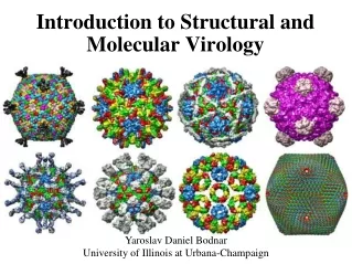
Physical Methods • The physical properties of viruses can be determined by, spectroscopy using either ultraviolet light to examine the nucleic acid content of the particle or visible light to determine its light-scattering properties. • Electrophoresis of intact virus particles has yielded some limited information, but electrophoretic analysis of individual virion proteins by gel electrophoresis, of nucleic acid genomes, has been far more valuable. • By far the most important method for the elucidation of virus structures has been the use of X-ray diffraction by crystalline forms of purified virus. This technique allows determination of the structure of virions at an atomic level. Joko Pamungkas
Chemical Methods • Classic studies of virus structure have been based on stepwise disruption of particles by slow alteration of pH, or gradual addition of protein-denaturing agents such as urea, phenol, or detergents. • 1)Proteins bound together by electrostatic interactions can be eluted by addition of ionic salts or alteration of pH, 2)those bound by non-ionic, hydrophobic interactions can be eluted by reagents such as urea, 3)proteins which interact with lipid components can be eluted by detergents or solvents. • Proteins exposed on the surface of viruses can be labelled with various compounds (e.g. iodine) to indicate which parts of the protein are exposed & which are protected inside the particle or by lipid membranes. Joko Pamungkas
Denaturation of TMV Joko Pamungkas
Electron Microscopy • Electron microscopes, developed in the 1930s, overcome the fundamental limitation of light microscopes, i.e. inability to resolve individual virus particles owing to physical constraints caused by the wavelength of visible light illumination & the optics of the instruments. • The first electron micrograph of a virus (TMV) was published in 1939. • During subsequent years, techniques were developed which allowed the direct examination of viruses at magnifications of over 100,000 times. • There are two fundamental types of electron microscope, the transmission electron microscope (TEM) & the scanning electron microscope (SEM). Joko Pamungkas
ElectronMicroscopy Joko Pamungkas
"Molecular Biology" • All the previous techniques of investigation are 'molecular biology' in the original sense. • However, the term 'molecular biology' (& 'genetic engineering' or 'genetic manipulation') has taken on a new & different meaning - manipulating nucleic acids in vitro. • Virus infection has long been used to probe the working of 'normal' (i.e. uninfected) cells, for example, to look at macromolecular synthesis. • Initially at least, the effect of this new technology was to shift the emphasis of investigation from proteins to nucleic acids. Joko Pamungkas
Hybridization Techniques • Nucleic acid-centred technology offers significant advances in detection of viruses & virus infections involving nucleic acid hybridization techniques. • There are many variants of this basic idea, but essentially, a hybridization probe, labelled in some fashion to facilitate detection, is allowed to react with a crude mixture of nucleic acids. • The specific interaction of the probe sequence with complementary virus-encoded sequences, to which it binds by hydrogen-bond formation between the complementary base pairs, reveals the presence of the virus genetic material. Joko Pamungkas