Download
1 / 83
900 likes | 935 Views
Complications of fractures. Done by: S elena abboud Rand A lshayeb Rahaf H asanain. COMPLICATIONS OF FRACTURES. General . Local . Early. Late. General complications. 1- Blood loss 2- Shock *Hypovolemic or hemorrhagic shock. *Septic shock.

E N D
Complications of fractures Done by: Selena abboud Rand Alshayeb RahafHasanain
COMPLICATIONS OF FRACTURES General Local Early Late
General complications • 1- Blood loss • 2- Shock • *Hypovolemic or hemorrhagic shock. • *Septic shock. • *Neurogenic shock. • 3- Fat embolism. • 4- Pulmonary embolism. • 5- Crush syndrome. • 6- Multiple organs failure syndrome (MOFS). • 7- Thrombo-embolism.
shock • follows a mismatch of metabolic demand to oxygen delivery at tissue level, leading to cellular hypoxia and (if uncorrected) to tissue and organ failure. • The causes of circulatory shock can be classified as abnormalities of cardiac output, of systemic vascular resistance, or a combination of both • HYPOVOLAEMIC SHOCK: Reduced circulating volume causing a reduction in venous return and cardiac output (e.g.haemorrhage) • Neurogenic shock : when spinal cord injury at a cervical or high thoracic level leads to loss of sympathetic tone and peripheral vasodilatation, venous pooling and reduced venous return.
*Management -ABC -Oxygen -fluids -Inotropes/vasopressors: if the patient remains hypotensive despite adequate fluid resuscitation -systemic support for other organs
Fat embolism • Circulating fat globules occlude small blood vessels, occur after closed fractures of long bones and traces of fat can be found in the lungs and other internal organs. • Source : bone marrow • more common in patients with multiple fractures. *Clinical features • Early warning signs of fat embolism (usually within 72 hours of injury) are 1- slight rise of temperature and pulse rate. 2- breathlessness ,mental confusion ,restlessness. 3- Pathognomonic signs are petechiae on the trunk, neck, axillae and conjunctiva. 4- Severe cases: respiratory distress and coma, due to brain emboli and hypoxia from involvement of the lungs. The features at this stage are essentially those of ARDS.
ABG and Urinalysis • There is no test for fat embolism • Management -supportive -oxygen -stabilization of long-bone fractures -analgesia -corticosteroids for severe multiple injuries(methylpredinosolone to maintain oxygen tension) -heparin or dextran ( improve capillary flow)
Crush syndrome • when a limb is compressed for extended periods – underperfusion - myonecrosis - release of toxic metabolites – reperfusion injury - Membrane damage and capillary fluid reabsorption failure – swelling - may lead to compartment syndrome - more tissue damage from ischaemia. • The compromised limb is pulseless,red, swollen,blistered; sensation and muscle power may be lost. • prevention. • high urine flow is encouraged with alkalization of the urine with sodium bicarbonate, prevents myoglobin precipitating in the renal tubules. • If compartment syndrome develops, and is confirmed by pressure measurements, then a fasciotomy is indicated. Excision of dead muscle must be radical to avoid sepsis. (( open wound then this should be managed)) . (( no open wound and compartment pressures are not high,risk of infection is lower if early surgery is avoided))
Multiple organ failure syndrome • (MODS) is the clinical appearance of a poorly controlled severe systemic inflammatory reaction, following a triggering event such as infection,inflammationor trauma. • the classical form of MODS appears to progress through four clinical phases: 1. Shock (hypoperfusion). 2. Period of active resuscitation. 3. Stable hypermetabolism (systemic inflammatory response). 4. Organ failure • Management: Prevention, remove risk factors, oxygen,treat underlying cause
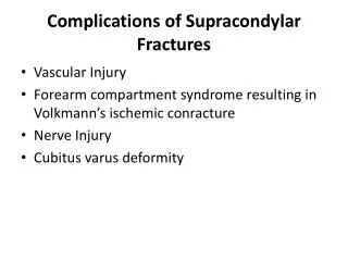
Local complications - Early • Visceral injury • Vascular injury. • Nerve injury. • Compartment syndrome. • Haemoarthrosis. • Infection. • Gas gangrene. • Fracture blisters. • Plaster and pressure sores
visceral Injury * Fractures around the trunk are often complicated by injuries to underlying viscera. 1) rib fractures : pneumothorax - penetration of the lung Spleen trauma Liver injury 2) pelvic fractures - rupture of the bladder or urethra. *These injuries require emergency treatment.
Vascular injury * The fractures most often associated with damage to a major artery are those around the knee and elbow, and those of the humeral and femoral shafts. * The artery may be cut, torn, compressed or contused either by the initial injury or subsequently by jagged bone fragments. * Even if its outward appearance is normal, the intima may be detached and the vessel blocked by thrombus, or a segment of artery may be in spasm. *The effects vary -transient diminution of blood flow -profound ischaemia -tissue death -peripheral gangrene.
InjuryVessel First rib fracture/clavicle Subclavian artery Shoulder dislocation Axillary artery Humeral supracondylar fracture Brachial artery Elbow dislocation Brachial artery Pelvic fracture Presacral and internal iliac artery Femoral supracondylar fracture Femoral artery Knee dislocation Popliteal artery Proximal tibial Popliteal artery or its branches Temporal/ parietal middle meningeal artery
Clinical features • - paraesthesia or numbness in the toes or the fingers. • -The injured limb is cold, pale, or slightly cyanosed. • -pulse is weak or absent. • -paralysis • -pain • If a vascular injury is suspected an angiogram should be performed immediately; if it is positive, emergency treatment must be started without further delay • Treatment • All bandages/splints removed • The fracture re X-Rayed • Circulation reassessed for next half hour • If no improvement, do vessels exploration • Suture torn vessels, vein grafting, if thrombosed do endarterectomy • Aim: to restore blood flow
Nerve Injury * Nerve injury is particularly common with fractures of the humerus or injuries around the elbow or the knee. Closed nerve injuries -The nerve is seldom severed. -spontaneous recovery awaited – occurs in 90% within 4 months. If no recovery by the expected time, and if nerve conduction studies fail to show evidence of recovery, the nerve should be explored. - By sharp edge of a bone Open nerve injuries -Nerve injury is more likely to be complete. -The nerve should be explored at the time of debridement and repaired at the time or at wound closure. -by agent causing fracture Acute nerve compression -Distinct from a direct injury, with fractures or dislocations around the wrist. Complaints of numbness or paraesthesia in the distribution of the median or ulnar nerves should be taken seriously and the patient monitored ; if there is no improvement within 48 hours of fracture reduction or splitting of bandages around the splint, nerve should be explored and decompressed
Compartment Syndrome • Fractures of the arm or leg can cause ischaemia, even if there is no damage to a major vessel. • Bleeding, oedema or inflammation (infection) may increase the pressure within one of the osseofascial compartments- reduced capillary flow- muscle ischaemia– oedema- greater pressure - more profound ischaemia • swelling of a limb inside a tight plaster cast. • – a vicious circle that ends after 12 hours or less, in necrosis of nerve and muscle within the compartment. • Nerve is capable of regeneration but muscle, once infarcted, can never recover and is replaced by inelastic fibrous tissue (Volkmann’s ischaemic contracture). • High-risk injuries are fractures of the • 1- elbow • 2- forearm bones • 3- proximal third of the tibia • 4- multiple fractures of the hand or foot • 5- crush injuries and circumferential burns. • 6- operation (usually for internal fixation) • 7- infection
The earliest of the ‘classic’ features are pain ( ‘bursting’ sensation), alteredsensibility and paresis (weakness in active muscle contraction). -Skin sensation repeatedly checked- • Ischaemic muscle is highly sensitive to stretch. (pain) • When the toes or fingers are passively hyperextended, there is increased pain in the calf or forearm. • Confirmation of the diagnosis is by measuring the intra compartmental pressures. • A differential pressure (ΔP) – the difference between diastolic pressure and compartment pressure – of less than 30 mmHg is an indication for immediate compartment decompression • A split/ wick catheter
Nerve damage may result in motor and sensory loss. • In extreme case gangrene Treatment -Decompression of the compartment. - Casts, bandages and dressings must be completely removed. -The limb should be nursed flat (elevating the limb causes a further decrease in end capillary pressure and aggravates the muscle ischaemia). -The ΔP monitored; if it falls below 30 mmHg, immediate open fasciotomy is performed. In the case of the leg, ‘fasciotomy’ means opening all four compartments through medial and lateral incisions. The wounds should be left open and inspected 2 days later: if there is muscle necrosis, debridement can be carried out; if the tissues are healthy, the wounds can be sutured (without tension) or skin-grafted. • if the clinical signs are ‘soft’, the limb should be examined at 30-minute intervals and if there is no improvement within 2 hours of splitting the dressings, fasciotomy should be performed. • Muscle will be dead after 4–6 hours of total ischaemia.
Haemarthrosis • Fractures involving a joint, leads to accumulation of blood. • (features) The joint is swollen and tense and the patient resists any attempt at moving it. The blood should be aspirated before dealing with the fracture.
Infection • Open fractures may become infected; closed fractures hardly ever do unless they are opened by operation. • Post-traumatic wound infection is now the most common cause of chronic osteitis. • Wound becomes inflamed and starts draining seropurulent fluid. • Infection may be superficial, moderate (osteomyelitis), severe (gas gangrene).
Treatment: • -Antibiotic • -Excising all devitalized tissue • -If there is pus formation : tissue around the fracture should be opened & drained
Gas gangrene especially Clostridium welchii • Produced by anaerobic organisms : Clostridium sp infections. • These organisms can survive and multiply in ↓ O2 tension • Toxins produced will destroy the cell wall and leads to tissue necrosis • Clinical features: within 24hr. Patient complains from : - intense pain - swelling around the wound - brownish discharge - gas formation - little or no pyrexia - characteristic smelling - toxaemic coma death • the prime site for infection is a dirty wound with dead muscle that has been closed without adequate debridement
Gas gangrene---characterized by myonecrosis • Anaerobic cellulitis in which superficial gas formation is abundant but toxaemia usually is slight. • Failure to recognize the difference may lead to • unnecessary amputation for the non-lethal cellulitis
swelling around the wound, brownish discharge gas formation
Prevention Deep, penetrating wounds in muscular tissue are dangerous; they should be explored, all dead tissue should be completely excised and, if there is the slightest doubt about tissue viability, the wound should be left open. There is no effective antitoxin against C. welchii. Treatment -early diagnosis. -fluid replacement -intravenous antibiotics -Hyperbaric oxygen has been used for limiting the spread of gangrene. -mainstay of treatment is prompt decompression of the wound and removal of all dead tissue. In advanced cases, amputation may be essential
Fracture blisters • Two distinct blister types are seen after fractures: • clear fluid-filled vesicles and blood-stained ones. • Both occur during limb swelling and are due to elevation of the epidermal layer of skin from the dermis . • There is no advantage to puncturing the blisters (it may even lead to increased local infection) and surgical incisions through blisters, whilst generally safe, should be undertaken only when limb swelling has decreased
Plaster and pressure sores • Plaster sores occur where skin presses directly onto bone. • They should be prevented by padding the bony points and by moulding the wet plaster so that pressure is distributed to the soft tissues around the bony points. • While a plaster sore is developing the patient feels localized burning pain. A window must immediately be cut in the plaster, or warning pain quickly abates and skin necrosis proceeds unnoticed. • careless selection of ring size, excessive fixed traction, and neglect can lead to pressure sores around the groin and iliac crest
Late complications • 1-DELAYED UNION • 2-NON-UNION • 3-MALUNION • 4-AVASCULAR NECROSIS • 5-GROWTH DISTURBANCE • 6-BED SORES • 7-MYOSITIS OSSIFICANS • 8-TENDON LESIONS • 9-NERVE COMPRESSION • 10-MUSCLE CONTRACTURE • 11-JOINT INSTABILITY • 12-JOINT STIFFNESS • 13-COMPLEX REGIONAL PAIN SYNDROME (ALGODYSTROPHY) • 14-OSTEOARTHRITIS
DELAYED UNION • If the time in which a fracture may be expected to unite and consolidateis prolonged, the term ‘ delayed union ’ is used .It must never be relied upon in deciding when treatment may be discontinued . • As a general rule , union is deemed to be delayed if the fracture is still freely mobile after 3 or 4 months . If a state of delayed union persists for many months it will eventually pass into a state of non-union • Causes can be summarized as :- 1- biological 2- biomechanical 3- patient-related
1- Biological cause :- A-Inadequate blood supply A badly displaced fracture of along bone will cause tearing of both the periosteum and interruption of the intramedullary blood supply . The fracture edges will become necrotic and dependent on the formation of an ensheathingcallus mass to bridge the break. If the zone of necrosis is extensive, as might occur in highly comminuted fractures, union may be interrupted . B-Severe soft tissue damage Severe damage to the soft tissues affects fracture healing by: (1) reducing the effectiveness of muscle splintage ; (2) damaging the local blood supply and (3) diminishing or eliminating the osteogenic input from mesenchymal stem cells within muscle. • C- Periosteal stripping over-enthusiastic stripping of periosteum during internal fixation is an avoidable cause of delayed union .
2- Biomechanical cause :- A-Imperfect splintage. Excessive traction (creating a fracture gap) or excessive movement at the fracture site will delay ossification in the callus. In the forearm and leg a single-bone fracture may be held apart by an intact fellow bone. B-Over-rigid fixation. Contrary to popular belief, rigid fixation delays rather than promotes fracture union. It is only because the fixation device holds the fragments so securely that the fracture seems to be ‘uniting’. Union by primary bone healing is slow, but provided stability is maintained throughout, it does eventually occur. C-Infection. Both biology and stability are affected by active infection: not only is there bone lysis, necrosis and pus formation, but implants which are used to hold the fracture tend to loosen.
3- Patient related :- There are patients who are :- • Immovable. • Impossible. • Age . • Associated systemic illness . • Smoking .
Clinical features :- • Fracture tenderness persists and, if the bone is subjected to stress, pain may be acute . • On x-ray, the fracture line remains visible and there is very little or incomplete callus formation or periosteal reaction. However, the bone ends are notsclerosed nor atrophic. The appearances suggest that , although the fracture has not united, it eventually will.
Treatment :- • CONSERVATIVE or OPERATIVE 1- conservative : • The two important principles are: (1) to eliminate any possible cause of delayed union and (2) to promote healing by providing the most appropriate environment. Immobilization (whether by cast or by internal fixation) should be sufficient to prevent shear at the fracture site, but fracture loading is an important stimulus to union and can be enhanced by: (1) encouraging muscular exercise and (2) by weightbearingin the cast or brace ?. The watchword is patience; however ,there comes a point with every fracture where the ill-effects of prolonged immobilization outweigh the advantages of non-operative treatment.
2- Operative :- • each case should be treated on its merits; however, if union is delayed for more than 6 months and there is no sign of callus formation, internal fixation and bone grafting are indicated. The operation should be planned in such a way as to cause the least possible damage to the soft tissues.
NON-UNION • In a minority of cases delayed union gradually turns into non-union that is it becomes apparent that the fracture will never unite without intervention. • Causes:- • When dealing with the problem of non-union, four questions must be addressed. They have given rise to the acronym CASS: • 1. Contact– Was there sufficient contact between the fragments? • 2. Alignment – Was the fracture adequately aligned , to reduce shear? • 3. Stability – Was the fracture held with sufficient stability? • 4. Stimulation – Was the fracture sufficiently ‘stimulated’? (e.g. by encouraging weightbearing).
There are, of course, also biological and patient-related reasons that may lead to non-union: • (1) distraction and separation of the fragments, sometimes the result of interposition of soft tissues between the fragments (there is muscle or ligament covering the broken ends and preventing them from touching each other) • (2) excessive movement at the fracture line (the two ends are still mobile ) . • (3) poor soft tissues (from either the injury or surgery) . • (4) poor local blood supply ( avascular necrosis ) . • (5) infection (particularly osteomyelitis ) . • (6) associated drug abuse, anti-inflammatory or cytotoxic immunosuppressant medication . • (7) non-compliance on the part of the patient . • (8) surgical intervention, If fill-judged, is another cause .
Clinical features :- • Movement can be elicited at the fracture site and pain diminishes ; the fracture gap ( cavity between the bone ends ) becomes a type of pseudoarthrosis. • X-ray The fracture is clearly visible but the bone on either side of it may show either exuberant callus or atrophy. This contrasting appearance has led to non-union being divided into hypertrophic and atrophic types. In hypertrophic non-union the bone ends are enlarged, suggesting that osteogenesis is still active but not quite capable of bridging the gap. In atrophic non-union, osteogenesis seems to have ceased. The bone ends are rounded with no suggestion of new bone formation.
Treatment :- • CONSERVATIVE or OPERATIVE 1- Conservative :- • Non-union is occasionally symptomless, needing no treatment as it might cause slight instability and is best left untreated ,or at most, (a) a removable splint. Even if symptoms are present, operation is not the only answer; with hypertrophic non-union, (b) functional bracing may be sufficient to induce union, but splintageoften needs to be prolonged. (c) Pulsed electromagnetic fields and low-frequency, pulsed ultrasoundcan also be used to stimulate union . • 2- Operative :- • More often , non-union of a fracture is disabling and surgical treatment is desirable , With hypertrophic non-union and in the absence of deformity, very rigid fixation alone (internal or external) may lead to union. With atrophic non-union, fixation alone is not enough. Fibrous tissue in the fracture gap, as well as the hard, sclerotic bone ends is excised and bone grafts are packed around the fracture. If there is significant ‘die-back’, this will require more extensive excision and the gap is then dealt with by bone advancement using the Ilizarov technique.
MALUNION • When the fragments join in an unsatisfactory position (unacceptable angulation, rotation or shortening) the fracture is said to be malunited. • Causes are • (a) failure to reduce a fracture adequately . • (b) failure to hold reduction while healing proceeds . • (c) gradual collapse of comminuted bone . • (d) osteoporotic bone .
Clinical features :- • The deformity is usually obvious, but sometimes the true extent of malunion is apparent only on x-ray . Rotational deformity of the femur, tibia, humerusor forearm may be missed unless the limb is compared with its opposite fellow. Rotational deformity of a metacarpal fracture is detected by asking the patient to flatten the fingers onto the palm and seeing whether the normal regular fan-shaped appearance is reproduced . • X-rays are essential to check the position of the fracture while it is uniting. This is particularly important during the first 3 weeks, when the situation may change without warning. At this stage it is sometimes difficult to decide what constitutes ‘malunion’ ; acceptable norms differ from one site to another .
Treatment :- • Incipient malunion may call for treatment even before the fracture has fully united; the decision on the need for re-manipulation or correction may be extremely difficult. A few guidelines are offered:- • 1. In adults, fractures should be reduced as near to the anatomical position as possible. Angulation of more than 10–15 degrees in a long bone or a noticeable rotationaldeformity may need correction by re-manipulation, or by osteotomy and fixation. • 2. In children, angular deformities near the bone ends (and especially if the deformity is in the same plane as that of movement of the nearby joint) will usually remodel with time; rotationaldeformities will not.
3. In the lower limb, shortening of more than 2.0 cm is seldom acceptable to the patient and a limb length equalizing procedure may be indicated. • *The patient’s expectations (often prompted by cosmesis) may be quite different from the surgeon’s; they are not to be ignored. • - Early discussion with the patient, and a guided view of the x-rays, will help in deciding the need for treatment and may prevent later misunderstanding. • - Very little is known of the long-term effects of small angular deformities on joint function . However, it seems likely that malalignment of more than 15 degrees in any plane may cause asymmetrical loading of the joint above or below and the late development of secondary osteoarthritis; this applies particularly to the large weightbearingjoints.
AVASCULAR NECROSIS • Death of a bone from a deficient blood supply . • Certain regions are known for their propensity to develop ischaemia and bone necrosis after injury . They are: • (1) the head of the femur (after fracture of the femoral neck or dislocation of the hip) • (2) the proximal part of the scaphoid (after fracture through its waist); • (3) the lunate (following dislocation) • (4) the body of the talus (after fracture of its neck). • Accurately speaking, this is an early complication of bone injury, because ischaemia occurs during the first few hours following fracture or dislocation. However , the clinical and radiological effects are not seen until weeks or even months later, the avascular bone gradually loses its rigid trabecular structure , in this state the bone crumbles easily and under stress imposed by the muscular tone or body weight it will eventually collapse which may be sometimes very slow.
Clinical features :- • Avascular necrosis causes : intractable non union , disabling osteoarthritis or total disorganization of a joint . • There are no symptoms associated with avascular necrosis, but if the fracture fails to unite or if the bone collapses the patient may complain of pain. • X-ray shows the characteristic increase in x-ray density , which occurs as a consequence of two factors: (a) disuse osteoporosis in the surrounding parts gives the impression of ‘increased density’ in the necrotic segment, and (b) collapse of trabeculae compacts the bone and increases its density. Where normal bone meets the necrotic segment a zone of increased radiographic density may be produced by new bone formation.