Download
1 / 26
530 likes | 1.09k Views
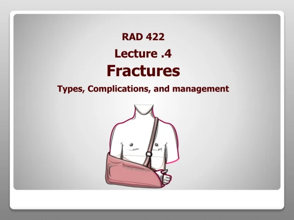
RAD 422 Fractures Types, Complications, and management. Lecture .4. L earning Objectives. With the end of the lecture the student should be able to:-. Define term the term fracture, dislocation and Subluxation Identify the general causes, signs , symptoms of fractures

E N D
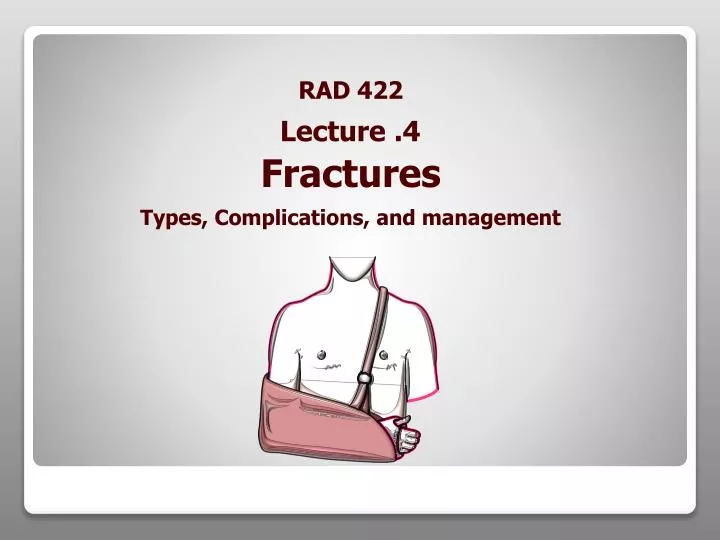
RAD 422 Fractures Types, Complications, and management Lecture .4
Learning Objectives With the end of the lecture the student should be able to:- • Define term the term fracture, dislocation and Subluxation • Identify the general causes, signs , symptoms of fractures • Classify the different types of fractures • Discus the general principles of the management of fractures • Describe the role of radiography in the management of fractures • Explain the process of fracture healing • State the local and general complications of fractures
References • Text book of radiographic positioning and related anatomy; by Kenneth • L.Bontrager,5th edition • M.A.Seraj , The new manual of basic emergency • procedures, first aid and updated CPR. Useful websites • http://www.medicinenet.com/fracture/article.htm • http://www.gentili.net/fracturemain.asp
What is fracture(#)? common causes of fractures: • Fall from a height • car accidents • Direct blow • Repetitive forces • Pathology a break in the continuity of a bone or cartilage. • Signs andSymptoms • Swelling or tenderness • pain • Numbness • Bleeding • Broken skin with bone protruding • Limitation or unwillingness to move a limb
Fracture Types 1/ Traumatic Closed fracture: A closed fracture is one where the fracture hematoma does not communicate with the outside Open fracture: This is one where the fracture hematoma communicates with the outside through an open wound. Stress fracture : It is a fracture occurring at a site in the bone subject to repeated minor stresses over a period of time. Birth fracture: It is a fracture in the new born children due to injury during delivery.
Fracture Types 2/ Pathological It is a fracture occurring after a trivial violence in a bone weakened by some pathological lesion. This lesion may be : - Localized disorder (e.g. secondary malignant deposit) - Generalized disorder (e.g. osteoporosis).
Fracture Types According to the Path of the # Line Oblique Fracture A fracture in which the # line is at oblique angle to the long axis of the bone. Transverse Fracture A fracture in which the # line is perpendicular to the long axis of the bone .
Fracture Types According to the Path of the # Line Longitudinal Fracture A fracture in which the # line runs nearly parallel to the long axis of the bone. A longitudinal fracture can be considered a long oblique fracture. Spiral Fracture A severe form of oblique fracture in which the # plane rotates along the long axis of the bone. These #s occur secondary to rotational force.
Fractures Anatomical classification of fractures Stellate fracture: This # occurs in the flat bones of the skull and in the patella, where the fracture lines run in various directions from one point. Comminuted # : The bone is broken into than two fragments.
Fracture Types Anatomical classification of fractures Impacted fracture: This # where a vertical force drives the distal fragment of the fracture into the proximal fragment. Depressed fracture: This # occurs in the skull where a segment of bone gets depressed into the cranium.
Fracture Types Anatomical classification of fractures Avulsion fracture: This is one, where a chip of bone is avulsed by the sudden and unexpected contraction of a powerful muscle from its point of insertion, Examples 1. The supra spinatus muscle avulsing the greater tuberosity of the humerus. 2. Avulsion fracture of the tibial tuberosity
Fracture Types Summary Simplelittle or no bone displacement Compound fracture ruptures the skin & bone protrudes Green stickoccurs mostly in children whose bones have not calcified or hardened Transverse crack perpendicular to long axis of the bone - displacement may occur Oblique diagonal crack across the long axis of the bone Spiral diagonal crack involving a "twisting" of the bone about the longitudinal axis Comminuted "crushing" fracture - more common in elderly Impacted one end of bone is driven up into the other Depressed broken bone is pressed inward (skull fracture) Avulsion fragment of bone is pulled away by tendon
Other Terms used in describing fracture Greenstick is the fracture in the young bone of children where the break is incomplete, leaving one cortex intact . Plastic - Bowing fracture in children without disruption of cortex. Distraction Is a separation of fragments that have been pulled apart. Distraction # Greenstick #
Terms used in fracture follow-up 1.Position - changed or unchanged .2. Healing -central or peripheral bony bridging
Terms used in fracture follow-up Delayed union - the healing process is slower than normal. Non-union - the healing stopped before union occurred. Malunion - the fracture healed in unacceptable position.
Principles of Management: Aims : (A)- safe life (B)-save the limb (C)-save the function Efficient First Aid: This relieves the pain and prevents complications. Safe transport:This help to minimize complications in injures to the spine, fracture of the lower limbs, ribs etc (all fractures should be immobilized immediately ) . Assessment of conditionof the patients for shock & other injuries. Assessment of local condition of the injured limb regarding complications like vascular injury, nerve involvement and injury to neighboring joints . Resuscitation. If needed Radiographyof the part • X-ray before plaster AP & LAT( to determine site and degree of displacement) • Post Reduction films ( wet plaster) for insurance of good alignment • Follow up films to assess healing • Films Before removal of plaster to confirm complete healing • 7. Reductionof the fracture(correction of displacement of fragments and done by : • closed Manipulation • open reduction
Principles of Management: • 8. Immobilization of the fragments. • External fixation • Cast (plaster) • Internal fixation • Screws • Plates • intramedullary nails and rod • wires & pins
Fractures are treated by reduction (realignment) &immediate immobilization • In most cases, simple fractures heal completely in approximately 6 - 8 weeks • Compound # better to deal with it within6hrs of injury to avoid infection • The accurate diagnosis of the fracture (site ,lines and displacement ) is made from X- ray examination. • Tow projections is required AP or PA +lateral or oblique • Tow joints above and below the site of the # should be included in the radiographs • Tow limbs radiographs for comparison of value in children. 9. Early physiotherapy : for the preservation of function of the limb (local complication such as ischemia ,nerve damage ,joint stiffness ,infection ..etc may endanger the function of the limb.10. Rehabilitation : After union of the fracture to restore full muscle power and joint movements and to make the patient fit for his original job.NOTE:
FRACTURE HEALING Fracture healing is considered as a series of phases which occur in sequence as follows: • (I) Inflammatory Phase. • Stage or hematoma formation. • Stage of granulation tissue. (more fibrin to the hematoma and increase blood flow • (II) Reparative Phase. • (A) Stage of fibro cartilaginous callus. • (B) Stage of bony callus (woven bone become calcified)). • (III) Remodeling Phase. • Excess material inside bone shaft is • replaced by more compact bone
At time of injury (Immediate) • Haemorrhage • Damage to important internal structures (brain ,heart..) • Skin loss ,Shock ,Nerve damage Later Complications Fracture - Complications
Joints Dislocation What is Dislocation? Is the total displacement of the articular end of a bone from the joint cavity. Subluxation : Is an incomplete displacement. Reduction : Is the restoration of the normal alignment of the bones. • Classification: • Dislocations are classified • as follows: • Congenital • Traumatic • Pathological • Paralytic
Dislocation & Subluxation (Sample Radiographs) Elbow joint Dislocation. PIPJ Subluxation
? U E S T Q I O S N Thank you