Download
1 / 60
650 likes | 823 Views
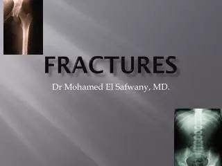
Fractures. Description. A disruption or break in the continuity of the structure of bone Traumatic injuries account for the majority of fractures. Description. Described and classified according to: Type Communication or noncommunication with external environment Anatomic location.

E N D
Description • A disruption or break in the continuity of the structure of bone • Traumatic injuries account for the majority of fractures
Description • Described and classified according to: • Type • Communication or noncommunication with external environment • Anatomic location
Types of Fractures Fig. 61-4
Classification by Communication withExternal Environment Fig. 61-5
Classification by Fracture Location Fig. 61-6
Description • Described and classified according to: • Appearance, position, and alignment of the fragments • Classic names • Stable or unstable
Description • Closed (also called simple) • Open (also called compound)
Description • Stable fractures • Occur when a piece of the periosteum is intact across the fracture • External or internal fixation has rendered the fragments stationary
Description • Unstable fractures • Grossly displaced • Poor fixation
Clinical Manifestations • Immediate localized pain • Function • Inability to bear weight or use affected part • Guarding • May or may not see obvious bone deformity
Fracture Healing • Reparative process of self-healing (union) occurs in the following stages: • Fracture hematoma (d/t bleeding, edema) • Granulation tissue → osteoid (3 – 14 days post injury) • Callus formation (minerals deposited in osteoid)
Fracture Healing • Reparative process of self-healing (union) occurs in the following stages: • Ossification (3 wks – 6 mos) • Consolidation (distance between fragments decreases → closes). • Remodeling (union completed; remodels to original shape, strength)
Bone Healing Fig. 61-7
Collaborative Care • Overall goals of treatment: • Anatomic realignment of bone fragments (reduction) • Immobilization to maintain alignment (fixation) • Restoration of normal function
Collaborative CareFracture Reduction • Closed reduction • Nonsurgical, manual realignment • Open reduction • Correction of bone alignment through a surgical incision
Collaborative CareFracture Reduction • Traction (with simultaneous counter-traction) • Application of pulling force to attain realignment • Skin traction (short-term: 48-72 hrs) • Skeletal traction (longer periods) • See Table 61-7
Collaborative CareFracture Immobilization • Casts • Temporary circumferential immobilization device • Common following closed reduction
Casts Fig. 61-9
Collaborative CareFracture Immobilization • External fixation • Metallic device composed of pins that are inserted into the bone and attached to external rods
Collaborative CareFracture Immobilization • Internal fixation • Pins, plates, intramedullary rods, and screws • Surgically inserted at the time of realignment
Collaborative CareFracture Immobilization • Traction • Application of a pulling force to an injured part of the body while countertraction pulls in the opposite direction
Collaborative CareFracture Immobilization • Purpose of traction: • Prevent or reduce muscle spasm • Immobilization • Reduction • Treat a pathologic condition
Nursing Management Nursing Assessment for Fractures • Brief history of the accident • Mechanism of injury • Special emphasis focused on the region distal to the site of injury
Nursing Management Nursing Assessment • Neurovascular assessment • Color and temperature • cyanotic and cool/cold: arterial insufficiency • Blue and warm: venous insufficiency • Capillary refill (want < 3 sec) • Peripheral pulses (↓ indicates vascular insufficiency)
Nursing Management Nursing Assessment • Neurovascular assessment • Edema • Sensation • Motor function • Pain
Nursing Management Nursing Diagnoses • Risk for peripheral neurovascular dysfunction • Acute pain • Risk for infection
Nursing Management Nursing Diagnoses • Risk for impaired skin integrity • Impaired physical mobility • Ineffective therapeutic regimen management
Nursing Management Nursing Implementation • General post-op care • Assess dressings/casts for bleeding/drainage • Prevent complications of immobility • Measures to prevent constipation • Frequent position changes/ ambulate as permitted • ROM exercised of unaffected joints • Deep breathing • Isometric exercises • Trapeze bar if permitted
Nursing Management Nursing Implementation • Traction • Ensure: • No frayed ropes, loose knots • Ropes in pulley grooves • Pulley clamps fastened securely • Weights must hang freely • Appropriate body alignment • Inspect skin • Around slings • Around pins
Nursing Management Nursing Implementation: Cast care • Casts can cause neurovascular complications if • Too tight • Edematous • Frequent neurovascular checks • Ice and elevation during early phase • See Table 61-10
Complications of FracturesInfection • Open fractures and soft tissue injuries have incidence • Osteomyelitis can become chronic
Complications of FracturesInfection • Collaborative Care • Open fractures require aggressive surgical debridement • Post-op IV antibiotics for 3 to 7 days (prophylactic)
Complications of FracturesCompartment Syndrome • Condition in which elevated intracompartmental pressure within a confined myofascial compartment compromises the neurovascular function of tissues within that space • Causes capillary perfusion to be reduced below a level necessary for tissue viability
Complications of FracturesCompartment Syndrome • Two basic etiologies create compartment syndrome: • Decreased compartment size (dressings, splints, casts) • Increased compartment content (bleeding, edema)
Complications of FracturesCompartment Syndrome • Clinical Manifestations • Six Ps • Paresthesia (unrelieved by narcotics) • Pain (unrelieved by narcotics) • Pressure
Complications of FracturesCompartment Syndrome • Clinical Manifestations • Six Ps: • Pallor (loss of normal color, coolness) • Paralysis • Pulselessness (decreased/absent pulses)
Complications of FracturesCompartment Syndrome • Clinical Manifestations • Six Ps: • Patient may present with one or all of the six Ps • Compare extemities
Complications of FracturesCompartment Syndrome • Clinical Manifestations • Absence of peripheral pulse = ominous late sign • Myoglobinuria • Dark reddish-brown urine
Complications of FracturesCompartment Syndrome • Collaborative Care • Prompt, accurate diagnosis is critical • Early recognition is the key • Do not apply ice or elevate above heart level
Complications of FracturesCompartment Syndrome • Collaborative Care • Remove/loosen the bandage and bivalve the cast • Reduce traction weight • Surgical decompression (fasciotomy)
Complications of FracturesVenous Thrombosis • Veins of the lower extremities and pelvis are highly susceptible to thrombus formation after fracture, especially hip fracture
Complications of FracturesVenous Thrombosis • Precipitating factors: • Venous stasis caused by incorrectly applied casts or traction • Local pressure on a vein • Immobility • Prevent with anticoagulant medications
Complications of FracturesFat Embolism Syndrome (FES) • Characterized by the presence of fat globules in tissues and organs after a traumatic skeletal injury
Complications of FracturesFat Embolism Syndrome (FES) • Fractures that most often cause FES: • Long bones • Ribs • Tibia • Pelvis
Complications of FracturesFat Embolism Syndrome (FES) • Tissues most often affected: • Lungs • Brain • Heart • Kidneys • Skin
Complications of FracturesFat Embolism Syndrome (FES) • Clinical Manifestations • Usually occur 24-48 hours after injury • Interstitial pneumonitis • Produce symptoms of ARDS
Complications of FracturesFat Embolism Syndrome (FES) • Clinical Manifestations • Symptoms of ARDS: • Chest pain • Tachypnea • Cyanosis • PaO2
Complications of FracturesFat Embolism Syndrome (FES) • Clinical Manifestations • Symptoms of ARDS: • Dyspnea • Apprehension • Tachycardia
Complications of FracturesFat Embolism Syndrome (FES) • Clinical Manifestations • Rapid and acute course • Feeling of impending disaster • Patient may become comatose in a short time