Download
1 / 49
490 likes | 498 Views
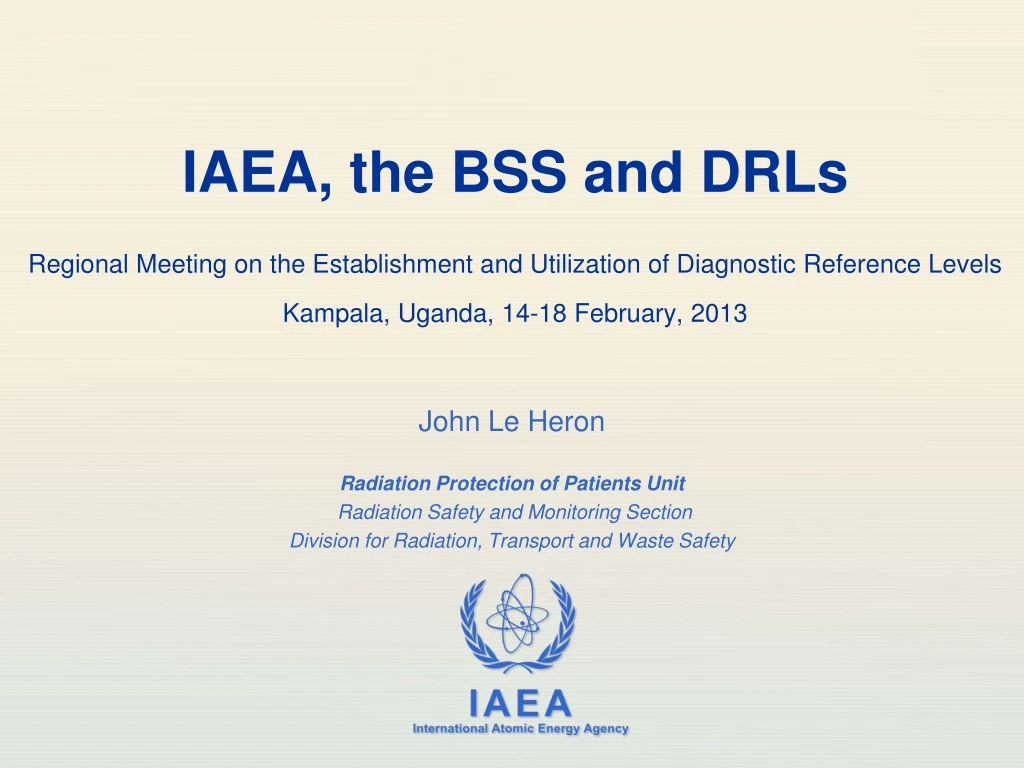
IAEA, the BSS and DRLs Regional Meeting on the Establishment and Utilization of Diagnostic Reference Levels Kampala, Uganda, 14-18 February, 2013. John Le Heron Radiation Protection of Patients Unit Radiation Safety and Monitoring Section Division for Radiation, Transport and Waste Safety.

E N D
IAEA, the BSS and DRLsRegional Meeting on the Establishment and Utilization of Diagnostic Reference LevelsKampala, Uganda, 14-18 February, 2013 John Le Heron Radiation Protection of Patients Unit Radiation Safety and Monitoring Section Division for Radiation, Transport and Waste Safety
Outline • Background & current issues • The BSS and radiation protection in medical exposures • IAEA activities & resources in TSA 3 • DRLs and the BSS
Medical exposures – current usage Every year, throughout the world, ionizing radiation is used in*: • 4.000.000.000 diagnostic procedures • 35.000.000 nuclear medicine procedures • 8.000.000 radiotherapy treatment courses • * An expanding activity worldwide These bring huge benefit to healthcare Diagnostic procedure Nuclear medicine procedure Radiotherapy procedure *UNSCEAR 2008
Increasing use of radiation in medicine • More machines, etc • New technologies and techniques • New roles • Increasing complexity in the planning & delivery of the radiation Single slice CT → Multi-Detector CT Film → Computed & Digital Radiography Hybrid imaging, PET-CT Image-guided interventional procedures Virtual procedures • E.g. Changes in the role of imaging: • First “port of call” • A move towards “screening”, in all its guises • E.g. IMRT, IGRT, etc.
Is this increasing use of radiation in medicine cause for concern? What are some of the current issues in imaging?
Whole body dose LD50 3000 - 5000 mSv X ray exams NBR, 2.4 mSv Patient doses – a perspective • Depends on the radiological procedure • E.g. Radiology: • Radiography • A few μSv to a few mSv, per procedure • CT • A few mSv to tens of mSv • Image-guided interventional procedures • A few mSv to tens of mSv • Skin doses up to several 1000 mSv • Radiation therapy • Many tens of Gy (but only to target vol)
Radiography • Doses to the patient are typically low • Effective dose – a few μSv to a few mSv • But variation by a factor of 20 more • Many exams lack proper justification and/or optimization
Image-Guided Interventional Procedures • Increase continues, in some countries doubling every 2 - 4 years • Doses can be high • Effective doses • Can exceed 20 mSv • Peak skin doses • Can exceed several Gy • Repeat procedures – not insignificant • Health professionals involved may not have had radiation protection training • Optimization often lacking
CT • Usage increasing • More scanners • Quicker to use • Can do more with them • But issues with: • Justification • Unnecessary exams • Self-referral • Pressure for “screening” • Optimization • Children • Multiple follow-up examinations
A need for radiation protection of the patient • ICRP principles of radiation protection • Justification • Net benefit for the patient • Optimization • Achieve clinical purpose with appropriate dose management Radiation dose Achieve clinical purpose
RP regulatory framework for medical exposure • The old BSS and the new BSS • The BSS sets out the requirements for Medical Exposure • Medical Exposure often called “TSA 3” in IAEA projects • Thematic Safety Area 3
IAEA projects and TSA 3 • Directed at end-users – medical radiation facilities • All hospitals and medical centres in a Member State where radiation is used in medical applications • i.e. From large teaching hospitals to small rural units • All modalities, as applicable • Diagnostic radiology • Radiography, fluoroscopy, CT, mammography, dental, DEXA • Image guided interventional procedures • Nuclear medicine • Radiation therapy
TSA 3 – for each medical radiation facility: • Appropriate persons are in place to take the relevant responsibilities • Radiological medical practitioners • Medical radiation technologists • Medical physicists
TSA 3 – for each medical radiation facility: • The radiation protection principle of justification is being applied • In particular, “Level 3” for individual justification
TSA 3 – for each medical radiation facility: • The principle of optimization of protection is being applied to every exposure • Design considerations for equipment • Operational considerations • Calibration • Dosimetry of patients • DRLs • Quality assurance for medical exposures • Dose constraints
TSA 3 – for each medical radiation facility: • Unintended and accidental medical exposures are being addressed • Means for minimizing their likelihood • If they occur: • Appropriate investigations • Appropriate corrective actions • Written records
Some IAEA activities to help Member States with radiation protection of the patient
Dedicated website – rpop.iaea.org Updated monthly Information for • Health professionals • Member States • Patients Additional resources • Publications • Safety Standards • Training material
The new BSS • Basis for RP in medical exposures • Safety Guide • RP in medical facilities (being developed) • Safety Report Series • Newer medical imaging techniques • Guidelines for the release of patients after radionuclide therapy • Establishing guidance levels in X ray guided medical interventional procedures
Promoting Education and Training • Development of standard packages for training in the application of the safety standards • Approved training packages on: • Radiation protection in: • Diagnostic and interventional radiology • Nuclear medicine • Radiotherapy • Cardiology • PET/CT • Paediatric radiology • Prevention of accidental exposure in radiotherapy • Dissemination of training material • Downloadable from RPoP website or available as CD • Organization of training courses
Technical Cooperation • Through regional and national projects: • Procurement for Member States • QC kits, phantoms, dosimeters, publications, etc • Fellowships & Scientific Visits • Expert missions • Regional & national training courses
The advent of DRLs Abdomen AP – NZ, 1983 • Large variations in patient doses for the same exam have been long documented • Many factors influence patient dose and image quality • The need for improvement long recognized • Various approaches advocated in 70s, 80s • E.g. Patient exposure guides (USA) • International recommendations • ICRP first mentioned “DRLs” in Publication 60, 1990 • Elaborated in Publication 73, 1996
The IAEA and DRLs • The International BSS, 1996 • Introduced Guidance Levels for medical exposure • Concept same as DRLs • Revised International BSS, 2011 • DRLs continue as an important tool for optimization of patient radiation protection in imaging
What does the new BSS require? • 2 aspects • Establishing (national) DRLs • Using the DRLs
Establishing national DRLs - BSS • Who? • Government as the facilitator • Health Authority • Professional Bodies • Regulatory Body
Establishing national DRLs - BSS • For what procedures? • Medical imaging • Including image guided interventional procedures
Using national DRLs - BSS • The (radiation protection) Regulatory Body mandates the use of the nationally established DRLs
Using national DRLs - BSS • At each medical radiation facility • Local assessments of typical doses for common procedures • Results compared with relevant DRLs, and if: • Exceed the relevant DRLs; or • Substantially below the relevant DRL and images not of diagnostic quality • Review of adequacy of optimization of patient radiation protection • Corrective action, if indicated
How DRLs work – a trigger for review • National DRLs have been established • Typical doses at a facility are periodically compared with the relevant DRLs • If exceeds DRL, or • If significantly below DRL and there are IQ problems • Investigate and if needed improve optimization DRL based on 75th percentile Average ESD Room AA = 4.4 mGy Average ESD Room BB = 6.9 mGy Note: if below DRL, still may not be optimized
What are the features of DRLs? • Applicable to a country or region within a country • Values established, in consultation, by • Professional bodies, Health Authority, RP Regulatory Body • For common examinations • In setting values, the following must be considered • Clinical requirements – general or specific • Adequate image quality • Use of easily measured dose quantities • Data from wide-spread surveys • Standardised patient or phantom • Need for revision as technology and techniques improve All of these will be discussed many times this week
In setting DRLs • Adequate image quality
Example – Image Quality & Mammography • 1990s, MGD increased • Image quality demands, including • Need for higher contrast • New film developed, higher density needed • Clinical requirements must be the driver • DRLs must not be an impediment to such developments * D Spelic, et al. Biomed Imaging and Interv 2007; 3(2):e35
Setting a DRL value for a procedure performed with different technologies and techniques
Example – Dental intra-oral radiography • Most common dental exam is the posterior “bitewing” view • Direct exposure film • D-speed • E/F-speed • Digital imaging • DR (mainly) • CR www.michigan.gov/mdch/0,1607,7-132-27417_35791_35798-46657--,00.htm M Alcaraz et al. Radiation Protection Dosimetry (2010) 140(4),391-5
Dental doses – intra-oral • Depends on the image receptor • Depends on the kVp, etc. • Factor of 5 in the example • Should the setting of DRLs accommodate all current practice or be technology specific? • National DRLs are based on wide-spread surveys • Blunt instrument • In parallel, the professional bodies must take the initiative • e.g. American Dental Association • Dentists should use E/F-speed film • In time, DRLs would reflect this professional body guidance http://www.michigan.gov/mdch/0,1607,7-132-27417_35791_35798-46657--,00.html
DRLs reflect immediate-past practice in a given country, “warts and all”, applied prospectively • Therefore, the periodic review of DRLs is very important
Patient size • The concept of a DRL is based on a typical patient, either: • A phantom, or • Patients selected on basis of some criteria • Does “looking after” this standardised patient ensure that all patients are ok? • Does an adult DRL help ensure optimization for a child? • Experience has shown that the answer is “No” • There is a need for a range of “standardised patients” • E.g. several paediatric sizes
Setting DRL values – not all exams are equal • DRLs for projection radiography are relatively easy • But with other modalities it is more difficult • Image Guided Interventional Procedures (IGIPs) • Factors include • Operator skill and experience • Patient size and anatomy • Complexity of the task • Equipment • Routine versus emergency • DRLs for IGIPs need to reflect the overall system • DRLs for IGIPs are not appropriate for deterministic effects • DRLs are not used for individual patients
DRL values and the new BSS • The new BSS gives no values • The old BSS did (Schedule III) • The new Safety Guide will discuss values of DRLs in use • Preference is for each country (or region in a country) to have their own • Based on the practice in their country
Do DRLs work – Trends with time • UK has > 20 years of experience with DRLs • Reviews in 1995, 2000, 2005 and 2010 • 2010 review showed for radiography: • On average about 16% lower than 2000 review • Typically less than 50% of original DRLs Trend due to better optimization, including regular monitoring of patient doses HPA-CRCE-034, Health Protection Agency, UK, 2012
Implementation around the world • Still a long way to go • Many countries have introduced DRLs, but the level of utilization varies widely • Between countries, and within countries • IAEA regional projects in patient protection • Developing Member States in: • Africa, Asia, Europe, Latin America • Includes setting up DRLs • Level of achievement to date is low • At RAF9044 RCM, DRLs were identified as the number 1 priority
Regional meeting, Kampala, 18-22 Feb • Aim • To describe, using teaching, practical work and group discussion, the concepts and methodologies that will enable participants to facilitate in their own countries the: • Establishment (and periodic review) of national DRLs, and • Application and use of DRLs in their country’s hospitals
Summary • BSS sets out the requirements for patient radiation protection • Optimization of protection is a cornerstone of patient radiation protection • DRLs are an important tool for optimization • Need to be established • Need to be used • Need to be reviewed periodically