Download
1 / 41
510 likes | 858 Views
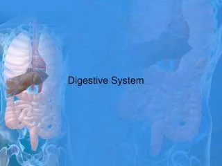
A Digestive Journey: From Food to Poop. Why do we need to eat?. Food provides us with : fuel to live, energy to work & play, and the raw materials to build new cells. 4 Components of Digestion. Ingestion – the taking of nutrients

E N D
Why do we need to eat? • Food provides us with: • fuel to live, • energy to work & play, and • the raw materials to build newcells.
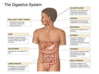
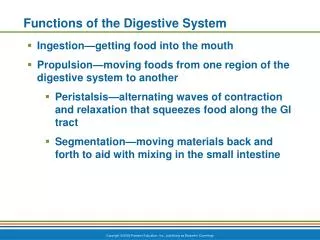
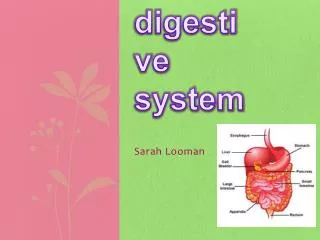
4 Components of Digestion Ingestion – the taking of nutrients • Digestion – the breakdown of complex organic molecules into smaller components by enzymes • Absorption – the transport of digested nutrients to the tissues of the body • Egestion – the removal of waste food materials from the body
A B C D
Ingestion A a c b d
Teeth • For Mechanical Breakdown • Incisors – for cutting • Canines – specialized for tearing • Premolars – specialized for grinding • Molars – for crushing
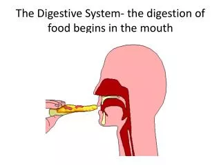
Tongue • Assists with the mechanical breakdown of food by pushing the food aroundwhile you chew with your teeth. • When you're ready to swallow, the tongue pushes a tiny bit of mushed-up food called a bolus (bow-lus) toward the back of your throat and into the opening of your esophagus.
Salivary Glands • Saliva (suh-lye-vuh) is produced in and secreted from salivary glands. • Chewing mixes the food with watery saliva, from 6 salivary glands around the mouth and face, to make it moist & slippery so it is mushy and easy to swallow. • Chemical breakdown of starch by production of salivary amylase from the salivary glands, an enzyme that begins the breakdown of starch into glucose. Functions of Saliva
Functions of Saliva • Lubrication &Binding: binding masticated food into a slippery bolus and coats the oral cavity and esophagus • Solubilizes Dry Food: in order to be tasted, the molecules in food must be solubilized • Oral Hygiene: oral cavity is almost constantly flushed with saliva, which floats away food debris and keeps the mouth relatively clean • Initiates Starch Digestion: amylase begins the breakdown of starch into glucose
Esophagus • The esophagus (ih-sah-fuh-gus) is a muscular tube whose muscular contractions (peristalsis) propel food from the back of your throat to the stomach. • But also at the back of your throat is your windpipe, which allows air to come in and out of your body. When you swallow a small ball of mushed-up food or liquids, a special flap called the epiglottis (eh-pih-glah-tiss) flops down over the opening of your windpipe to make sure the food enters the esophagus and not the windpipe.
Digestion B d a e b c
Stomach • It's a stretchy sack shaped like the letter J with thick muscles in its wall that contract to mash the food. • The stomach walls also release gastric(gas-trik) juices that assists in the breaking down of food and also helps to kill bacteria that might be in the eaten food. • It has four basic functions that assist in the early stages of digestion and prepare the ingesta for further processing in the small intestine:
Functions of the Stomach • to store the food you've eaten, allowing a rather large meal to be consumed quickly and dealt with over an extended period of time; • to attack the food in a chemical way, breaking down and dissolving its nutrients, it is in the stomach that substantial chemical and enzymatic digestion is initiated; • to break down the food into a liquidy mixture through the vigorous contractions of gastric smooth muscle which mix and grind foodstuffs with gastric secretions, resulting in liquefaction of food, a prerequisite for delivery of the ingesta to the small intestine; and • to slowly empty that liquidy mixture into the small intestine, as food is liquefied in the stomach, it is slowly released into the small intestine for further processing.
Pancreas • The pancreas (pan-kree-us), like the stomach, makes powerful digestive juices called enzymes which help to digest food further, specifically fats & proteins (into small peptide fragments and some amino acids by the enzyme proteases), as it enters the small intestines. • The pancreas sends pancreatic juice, which neutralizes the chyme (the mix of acid and food in the stomach), to the small intestine through the pancreatic duct. continued …
Pancreas, continued ... • The pancreas plays a vital role in accomplishing both of the previousobjectives, so vital in fact that insufficient exocrine secretion by the pancreas leads to starvation, even if adequate quantities of high quality food is consumed. • In addition to its role as an exocrine organ, the pancreas is also an endocrine organ and the major hormones it secretes - insulin and glucagon - play a vital role in carbohydrate and lipid metabolism.
Small Intestine • The small intestine is the major site for digestion and absorption of nutrients. • It is a long tube that's about 3.5 to 5cm around, and it's packed beneath the stomach, if stretched out, an adult's small intestine would be about 6.7m long. • The upper part, the duodenum, is the most active in digestion, where the digestion of carbohydrates, proteins, and fats continues. continued …
Small Intestine, continued ... • Starch and glycogen are broken down into maltose. • Food may spend as long as 4 hours in the small intestine and it will become a very thin, watery mixture so the nutrients are small enough to pass through the lining of the small intestine, and into the blood, where they are carried away to the liver and other body parts to be processed, stored and distributed.
Liver • The liver is the largest gland in the body and performs an astonishingly large number of tasks that impact all body systems. • For digestion, the liver produces bile, which is stored in the gall bladder before entering the bile duct into the duodenum. • Bile emulsifies fats, facilitating their breakdown into progressively smaller fat globules until they can be acted upon by lipases.Fats are completely digested in the small intestine, unlike carbohydrates and proteins.
Gall Bladder • This small bag-like part is tucked under the liver. • It stores a fluid called bile, which is made in the liver. • As food from a meal arrives in the small intestine, bile flows from the gall bladder along the bile duct into the intestine. • It helps to digest fatty foods and also contains wastes for removal.
Absorption C a c b
Stomach • The stomach absorbs some water, specific vitamins, some medicines, & alcohol.
Small Intestine • The net effect of passage through the small intestine is absorption of most of the water and electrolytes (sodium, chloride, potassium) and essentially all dietary organic molecules (including glucose, amino acids and fatty acids). Through these activities, the small intestine not only provides nutrients to the body, but plays a critical role in water and acid-base balance. • Most absorption occurs in the duodenum and jejeunum (second third of the small intestine). More
Villi & Microvilli • The inner surface of the intestine has circular folds that more than triple the surface area for absorption. Villi covered with epithelial cells increase the surface area by another factor of 10. The epithelial cells are lined with microvilli that further increase the surface area; a 6m long tube has a surface area of 300 square metres. • Each villus has a surface that is adjacent to the inside of the small intestinal opening covered in microvilli that form on top of an epithelial cell known as a brush border. Each villus has a capillary network supplied by a small arteriole. Absorbed substances pass through the brush border into the capillary, usually by passive transport. More
Villi & Microvilli, continued … • Maltose, sucrose, and lactose are the main carbohydrates present in the small intestine; they are absorbed by the microvilli. Starch is broken down into two-glucose units (maltose) elsewhere. Enzymes in the cells convert these disaccharides into monosaccharides that then leave the cell and enter the capillary. • Peptide fragments and amino acids cross the epithelial cell membranes by active transport. Inside the cell they are broken into amino acids that then enter the capillary.
Large Intestine • The large intestine is made up by the colon (coh-lun), cecum, appendix, and rectum. • Material in the large intestine is mostly indigestible residue and liquid. • Water, salts, and vitamins are absorbed back into the blood, the remaining contents form feces. • Bacteria in the large intestine, such as E. coli, produce vitamins (including vitamin K) that are absorbed. • The part of the large intestine called the colon, is where the body gets its last chance to absorb the water and some minerals into the blood.
Egestion D a
Rectum & Anus • As the water leaves the waste product, what's left gets harder and harder as it keeps moving along, until it becomes a solid, ready to be removed from the body. • The large intestine pushes the poop into the rectum (rek-tum), the very last stop on the digestive tract. • The solid waste stays here until you are ready to go to the bathroom. When you go to the bathroom, you are getting rid of this solid waste by pushing it through the anus (ay-nus), a ring of muscle and out of the body.
Sources: http://www.emc.maricopa.edu/faculty/farabee/BIOBK/BioBookDIGEST.html http://kidshealth.org/kid/body/digest_noSW.html http://users.tpg.com.au/users/amcgann/body/digestive.html http://arbl.cvmbs.colostate.edu/hbooks/pathphys/digestion/ http://www.bupa.co.uk/health_information/html/organ/stomach.html http://www.nlm.nih.gov/medlineplus/ency/imagepages/19221.htm http://www.bupa.co.uk/health_information/html/organ/liver.html http://trms.sheridank12.net/New%20Homepage/Gen.www/Brian%20genY/Brian%20Images/gall_bladder.htm http://www.nlm.nih.gov/medlineplus/ency/imagepages/8832.htm http://vanderbiltowc.wellsource.com/dh/content.asp?ID=574