Download
1 / 13
130 likes | 236 Views
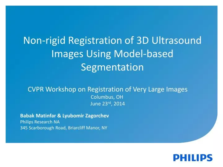
Non-rigid Registration of 3D Ultrasound Images Using Model-based Segmentation. Babak Matinfar & Lyubomir Zagorchev. Philips Research NA 345 Scarborough Road, Briarcliff Manor, NY. CVPR Workshop on Registration of Very Large Images Columbus, OH June 23 rd , 2014. Motivation.

E N D
Non-rigid Registration of 3D Ultrasound Images Using Model-based Segmentation Babak Matinfar & LyubomirZagorchev Philips Research NA 345 Scarborough Road, Briarcliff Manor, NY CVPR Workshop on Registration of Very Large Images Columbus, OH June 23rd, 2014
Motivation • Non-rigid registration computational performance depends on the image size • Voxel-based approaches are computationally intensive, exponentially proportional to image resolution • An approach that is independent of the image size • Registration computation speed remains constant with increasing image resolution
Method • Model based segmentation: • Fully automated, rapid and highly accurate segmentation on 3D volumes • Fixed mesh topology for point correspondence between segmented volumes • Thin plate spline (TPS) registration • TPS applied to segmented meshes from 4D echocardiogram images
4D Echocardiography Frame Extraction Frame Extraction Reference 3D ultrasound volume Target 3D ultrasound volume Model based segmentation Model based segmentation 3D mesh 3D mesh TPS Registration Control Points Control Points Registered reference and target images
3D ultrasound volumes were extracted from 4D echocardiography at different points in the cardiac cycle
Shape-constrained Deformable Models Training Anatomical knowledge Sample images Generic model Þ + Adaptation Generic model New image Adapted model Þ +
Segmentation of heart aortic root model Distance error
Results: Inter-Patient Registration Registered Not Registered Image overlay of end of diastole to end of systole volumes in cardiac cycle
Results: Inter-Patient Registration Not Registered Registered Overlay of image boundaries of end of diastole to end of systole volumes in cardiac cycle
Results: Intra-Patient Registration Registered Not Registered Image overlay of end of diastole volumes in different patients
Conclusion • Method for non-rigid registration of large 3D data volumes that does not depend on the image size and any intensity-based similarity metrics • Rapid segmentation provides accurate point-based correspondence that can be used for TPS registration • Registration computational performance remains constant with increasing image size and resolution