Download
1 / 22
220 likes | 662 Views
THE AUTONOMIC NERVOUS SYSTEM (ANS). What is the autonomic nervous system?. The autonomic nervous system (ANS) is the motor division of the peripheral nervous system that controls visceral activities, with the goal of maintaining internal homeostasis. What does the autonomic nervous system do?.

E N D
What is the autonomic nervous system? • The autonomic nervous system (ANS) is the motor division of the peripheral nervous system that controls visceral activities, with the goal of maintaining internal homeostasis.
What does the autonomic nervous system do? • It provides motor fibers to smooth and cardiac muscles and glands • It operates subconsciously • It causes excitation and inhibition • It makes adjustments to support body activities • It has an efferent pathway made up of a two-neuron chain: preganglionic and ganglionic
Two divisions of the ANS • 1) Parasympathetic division • 2) Sympathetic Although they have different roles, they have effects on many of the same organs of the body
Roles of the two different divisions 1. The parasympathetic division… _conserves body energy and maintains body activities at basal levels _is involved in digestion, diuresis, and defecation _causes heart rate and blood pressure to be low, the skin to be warm and the pupils to be constricted
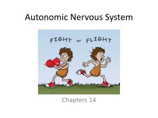
2. The sympathetic division… _activates the body under conditions of emergency which is why it is often called the “fight or flight system” _is involved in emergency, exercise, and excitement _causes blood flow to organs to reduce and blood flow to muscles to increase _also causes skin to be cold, heart rate to increase, and rapid breathing
Parasympathetic division outflow • Neurons of the cranial and sacral outflows of the parasympathetic division are located in the following nerves and create an effect on the organs mentioned in the table below.
Sympathetic division outflow Sympathetic division outflow is different from parasympathetic division outflow because… • It arises from spinal cord segments T1 through L2 • Preganglionic fibers pass through the white rami communicantes and synapse in the chain (paravertebral) ganglia • Fibers from T5-L2 form splanchnic nerves and synapse with collateral ganglia • Postganglionic fibers innervate the numerous organs of the body • Sympathetic neurons produce the lateral horns of the spinal cord
ANS PhysiologyNeurotransmitters and Receptors • The two major neurotransmitter involved in the ANS are: • Acetylcholine (ACh) • Norepinephrine (NE) • ACh is the same neurotransmitter that is found in the somatic motor neurons and is released in the ANS: • All ANS preganglionic axons • All parasympathetic postganglionic axons at synapses with their effectors • ACh releasing fibers are called Cholinergic Fibers • NE is released by most sympathetic postganglionic axons • NE releasing fibers are called Adrenergic Fibers • The effects of ACh and NE are either excitation or inhibition which is dependant on the receptor type allowing them to exert these different effects at different areas in the body
Receptors • ACh binds to two types of receptors: • Nicotinic • Muscarine • Nicotinic Receptors are located on: • Motor end plates of skeletal muscles (somatic targets) • All ganglionic neurons (sypathetic and parasympathetic) • The hormone-producing cells of the adrenal medulla • ACh always produces a stimulatory effect when it binds with nicotinic receptors • Muscarine Receptors occur on all effector cells stimulated by postganglionic cholinergic fibers (parasympathetic targets like the eccrine sweat glands and some blood vessels of skeletal muscle) • ACh binding produces a inhibitory or excitatory depending on the receptor type of the target organ
Receptors • There are also receptors know as Adrenergic Receptors (two types): • Alpha with two subtypes (A1,A2) • Beta with three subtypes (B1,B2,B3) • The general effects of NE binding are: • Alpha receptors are mostly stimulatory • Beta receptors are mostly inhibitory • However, an exception to this is when NE binds to the Beta receptors of cardiac muscle and the result is stimulatory
Drugs and the ANS • Knowing the locations of the cholinergic and adrenergic receptors subtypes allows specific drugs to be prescribed to obtain the desired inhibitory or stimulatory effect on selected organs • An example of this is Atropine (blocks parasympathetic effects): • administered during surgery to prevent salivation and dry up respiratory secretions • Ophthalmologists use it to dilate the pupils for an eye exam • There are several examples of drugs in Table 14.4 that influence the Activity of the ANS on page 544
Interactions of the Autonomic Divisions • Most visceral organs receive innervation by both sympathetic and parasympathtic fibers • This dual innervation produces a dynamic antagonism that allows visceral activity to be precisely controlled • Sympathetic fibers increase heart and respiratory rates. They also inhibit digestion and elimination • Parasympathetic fibers allow for digestion and elimination and decrease respiratory and heart rates
Parasympathetic & Sympathetic • The parasympathetic division is know as the “resting and digesting” division • The sympathetic division controls blood pressure and keeps the blood vessels in a continual state of partial constriction • Flight or Fright response – the sympathetic can override the parasympathetic in an emergency to increase heart and respiratory rates while inhibiting digestion. • Parasympathetic division restores the heart and respiratory rates back to resting levels when the emergency is over and then returns back to it’s functions of digestion and elimination
Sympathetic Tone& Parasympathetic Tone • The Sympathetic Tone: • Constricts blood vessels & controls blood pressure to rise in response to the body’s needs • Prompts vessels to dilate if blood pressure needs to be decreased • The Parasympathetic Tone: • Slows the heart • Controls Digestion and Elimination • ANS control is best seen in the external genitalia: • Parasympathetic fibers cause vasodilation resulting in the erection of the penis and clitoris • Sympathetic fibers cause ejaculation of sperm and reflex peristalsis in females
Sympathetic Division • The adrenal medulla, sweat glands and the arrector pili muscles of the skin, the kidneys, and most blood vessels receive only sympathetic fibers. • The Sympathetic Division controls: • Thermoregulatory Responses to heat • Release of Renin from the kidneys • Metabolic Effects
Control of Autonomic Functions Autonomic functions are controlled by several factors: • Reflex Activities mediated by the Spinal Cord and Brain Stem (Medullary Center) • Hypothalamic integration centers interact with both higher and lower centers of autonomic, somatic, and endocrine response • Cortical centers influence autonomic functioning via connections with the limbic system; conscious controls are rare but maybe learned via biofeedback training