Download
1 / 70
710 likes | 815 Views
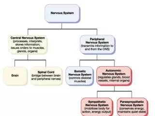
14 The Autonomic Nervous System. Section 1: ANS Functional Anatomy and Organization. Learning Outcomes 14.1 List the divisions of the ANS and the general functions of each.

E N D
14 The Autonomic Nervous System
Section 1: ANS Functional Anatomy and Organization Learning Outcomes 14.1 List the divisions of the ANS and the general functions of each. 14.2 Describe the structures and functions of the sympathetic and parasympathetic divisions of the autonomic nervous system. 14.3 Describe the innervation patterns of the sympathetic and parasympathetic divisions of the autonomic nervous system.
Section 1: ANS Functional Anatomy and Organization Learning Outcomes 14.4 Describe the various types of sympathetic and parasympathetic receptors and their associated neurotransmitters. 14.5 Describe the mechanisms of neurotransmitter release in the ANS, and explain the effects of neurotransmitters on target organs and tissues.
Section 1: ANS Functional Anatomy and Organization Comparisons of somatic nervous system (SNS) and autonomic nervous system (ANS) SNS Motor neurons exert voluntary control over skeletal muscles Lower motor neurons may be controlled by Reflexes based in spinal cord Upper motor neurons with cell bodies in brain nuclei or at primary motor cortex Animation: Somatic Autonomic Nervous System
Figure 14 Section 1 1 A schematic of the somatic nervous system (SNS), which provides conscious and sub- Conscious control over skeletal muscles Upper motor neurons in primary motor cortex BRAIN Somatic motor nuclei of brain stem Skeletal muscle Spinal cord Lower motor neurons Somatic motor nuclei of spinal cord Skeletal muscle
Section 1: ANS Functional Anatomy and Organization Comparisons of somatic nervous system (SNS) and autonomic nervous system (ANS) (continued) ANS CNS motor neurons synapse with visceral motor neurons in autonomic ganglia, which control visceral effectors Integrative centers located in hypothalamus Comparable to SNS upper motor neurons Two types of visceral motor neurons Preganglionic neurons (cell bodies in CNS) Activities represent direct reflex responses Ganglionic neurons (cell bodies in autonomic ganglia) Innervate effectors like cardiac and smooth muscle
Figure 14 Section 1 2 Visceral motor nuclei in hypothalamus BRAIN Preganglionic neuron Visceral Effectors Smooth muscle Autonomic nuclei in brain stem Autonomic ganglia Glands Ganglionic neurons Spinal cord Cardiac muscle Autonomic nuclei in spinal cord Adipocytes Preganglionic neuron A schematic of the autonomic nervous system (ANS), which controls visceral functions largely outside our awareness
Module 14.1: ANS divisions Three ANS divisions Sympathetic (or thoracolumbar) division Axons emerge from thoracic and superior lumbar segments of spinal cord Innervate ganglia relatively close to spinal cord “Kicks in” only during periods of exertion, stress, or emergency Parasympathetic (or craniosacral) division Axons emerge from brain stem and sacral spinal segments Innervate ganglia very close (or within) target organs Most often, effects are opposite to sympathetic
Figure 14.1 1 Autonomic Nervous System Sympathetic Division Parasympathetic Division In the sympathetic division, or thoracolumbar (thor-a-kō-LUM-bar) division, axons emerge from the thoracic and superior lumbar segments of the spinal cord and innervate ganglia relatively close to the spinal cord. In the parasympathetic division, or cranio- sacral (krā-nē-ō-SĀ-krul) divions, axons emerge from the brain stem and the sacral segments of the spinal cord, and they innervate ganglia very close to (or within) target organs. Cranial nerves (III, VII, IX, and X) T1 T2 T3 T4 The two main divisions of the ANS: the sympathetic and parasympathetic divisions T5 T6 Thoracic nerves T7 T8 T9 T10 T11 T12 L1 Lumbar nerves (L1, L2 only) L2 S2 Sacral nerves (S2, S3, S4 only) S3 S4
Module 14.1: ANS divisions Three ANS divisions (continued) Enteric nervous system (ENS) Extensive network of neurons (~100 million) and nerve networks within walls of digestive tract Influenced by sympathetic and parasympathetic divisions However, many complex visceral activities are coordinated on a local level (without CNS instructions) Will be discussed more in Ch. 21 (Digestive System)
Figure 14.1 2 The extensive system of neurons and nerve networks of the enteric nervous system (ENS) Esophagus Stomach Large intestine Small intestine
Figure 14.1 3 Aortic arch Right vagus nerve Trachea Autonomic Plexuses and Ganglia Left vagus nerve Cardiac plexus Thoracic spinal nerves Pulmonary plexus Esophagus Thoracic sympathetic chain ganglia Esophageal plexus Splanchnic nerves Celiac plexus and ganglion Superior mesenteric ganglion Diaphragm Superior mesenteric artery Inferior mesenteric plexus and ganglion Inferior mesenteric artery Hypogastric plexus Pelvic symapthetic chain Representative plexuses, ganglia, and nerves of the sympathetic and parasympathetic divisions
Module 14.1 Review a. Identify the major divisions of the autonomic nervous system. b. What division of the ANS is responsible for the physiological changes you experience when startled by a loud noise? c. Compare the anatomy of the sympathetic division with the parasympathetic division.
Module 14.2: Autonomic ganglia Autonomic ganglia Sympathetic division Preganglionic fibers (neurons) are relatively short while postganglionic fibers (neurons) are relatively long Accordingly, sympathetic ganglia (where these fibers synapse) are relatively near spinal cord Specific ganglia Sympathetic chain (on either side of spinal cord) Innervates visceral effectors in thoracic cavity, head, body wall, and limbs
Module 14.2: Autonomic ganglia Sympathetic division (continued) Specific ganglia (continued) 2.Collateral ganglia (within abdominopelvic cavity) Includes celiac, superior, and inferior mesenteric ganglia Innervates visceral effectors in abdominopelvic cavity Adrenal medulla Center of adrenal gland Acts as endocrine gland Targets organs and systems throughout body
Module 14.2: Autonomic ganglia Sympathetic division (continued) Prepares body for heightened levels of somatic activity Known as “fight or flight” division Typical responses Heightened mental alertness Increased metabolic rate Reduced digestive and urinary functions Activation of energy reserves Increased respiratory rate and dilation of passageways Elevated heart rate and blood pressure Activation of sweat glands
Figure 14.2 1 Organization of the sympathetic division of the ANS KEY Preganglionic fibers Postganglionic fibers Ganglionic neurons in the sympathetic chain and collateral ganglia exert their effects through innervation of peripheral target organs. Hormones released into circulation Target Organs Ganglionic Neurons Each sympathetic chain consists of a series of interconnected ganglia located on either side of the vertebral column. Visceral effectors in thoracic cavity, head, body wall, and limbs Preganglionic Neurons Lateral gray horns of spinal segments T1-L2 The collateral ganglia, located within the abdominopelvic cavity, include the celiac, superior mesenteric, and inferior mesenteric ganglia. Visceral effectors in abdomino- pelvic cavity Organs and systems throughout the body The center of each adrenal gland contains a sympathetic ganglion, the adrenal medulla, that acts as an endocrine organ. Ganglionic neurons in the adrenal medullae affect target organs throughout the body through the release of hormones into the general circulation.
Module 14.2: Autonomic ganglia Parasympathetic division Typical preganglionic fiber synapses on 6–8 ganglionic neurons May be situated in: Terminal ganglia (near target organ) Usually paired Ciliary ganglion (intrinsic eye muscles) Pterygopalatine and submandibular ganglia (nasal, tear, and salivary glands) Otic ganglion (parotid salivary gland)
Module 14.2: Autonomic ganglia Parasympathetic division (continued) Typical preganglionic fiber synapses on 6–8 ganglionic neurons (continued) May be situated in: (continued) Intramural (murus, wall) ganglia (embedded in target organ wall) Typically interconnected masses/clusters of cells Innervate visceral organs of neck, and of thoracic and abdominopelvic cavities
Module 14.2: Autonomic ganglia Parasympathetic division (continued) Concerned with regulation of visceral function and energy conservation Known as “rest and digest” system Typical responses Decreased metabolic rate Decreased heart rate and blood pressure Increased salivary and digestive gland secretion Increased digestive tract motility and blood flow Stimulation of urination and defecation
Figure 14.2 2 Organization of the parasympathetic division of the ANS KEY Preganglionic fibers Postganglionic fibers Preganglionic Neurons Ganglionic Neurons Target Organs III Intrinsic eye muscles (pupil and lens shape) Ciliary ganglion The midbrain, pons, and medulla oblongata contain parasympathetic nuclei associated with cranial nerves III, VII, IX, and X. VII Pterygopalatine and submandibular ganglia Nasal glands, tear glands, and salivary glands IX X Otic ganglion Parotid salivary gland Visceral organs of neck, thoracic cavity, and most of abdominal cavity Intramural ganglia In sacral segments of the spinal cord, parasympathetic nuclei lie in the lateral gray horns of spinal segments S2–S4. Visceral organs in inferior portion of abdominopelvic cavity Pelvic nerves Intramural ganglia
Module 14.2 Review a. List general responses to increased sympathetic activity and to parasympathetic activity. b. Describe an intramural ganglion. c. Starting in the spinal cord, trace the path of a nerve impulse through the sympathetic ANS to its target organ in the abdominopelvic cavity.
Module 14.3: Autonomic innervation patterns Autonomic innervation patterns Sympathetic division Every spinal nerve has a gray ramus carrying sympathetic postganglionic fibers Preganglionic fibers passing to collateral ganglia form splanchnic nerves Postganglionic fibers innervating thoracic cavity structures form bundles or sympathetic nerves
Figure 14.3 The innervation of the sympathetic division: at left, the distribution of nerves to the skin, skeletal muscles, and tissues of the body wall; at right, the distribution of nerves to visceral organs Eye PONS Salivary glands Sympathetic nerves Superior Cervical sympathetic ganglia Middle Heart Inferior Cardiac and pulmonary plexuses T1 Splanchnic nerves (SEE NOTE 2) T1 Gray rami to spinal nerves (SEE NOTE 1) Celiac ganglion Lung Superior mesenteric ganglion Liver and gallbladder Stomach Spleen Pancreas Large intestine Postganglionic fibers to spinal nerves (innervating skin, blood vessels, sweat glands, arrector pili muscles, adipose tissue) Small intestine Inferior mesenteric ganglion L2 L2 Adrenal medulla Spinal cord Sympathetic chain ganglia Kidney Ovary Coccygeal ganglia (Co1) fused together Penis Urinary bladder Uterus NOTE 1 Scrotum Every spinal nerve has a gray ramus that carries sympathetic postganglionic fibers for distribution in the body wall and limbs. In the head and neck, postganglionic sympathetic fibers leaving the superior cervical sympathetic ganglia supply the regions innervated by cranial nerves III, VII, IX, and X. NOTE 2 Preganglionic fibers on their way to the collateral ganglia form the splanchnic (SPLANK-nik) nerves. Postganglionic fibers innervating structures in the thoracic cavity, such as the heart and lungs, form bundles known as sympathetic nerves. KEY Preganglionic neurons Ganglionic neurons
Module 14.3: Autonomic innervation patterns Parasympathetic division Vagus nerve (X) alone provides ~75% of all parasympathetic outflow Numerous vagus nerve branches intermingle with sympathetic fibers forming nerve plexuses Preganglionic fibers in sacral spinal cord segments form distinct pelvic nerves Innervate intramural ganglia in kidneys, bladder, terminal portions of large intestine, and sex organs
Figure 14.3 The innervation of the parasympathetic division on one side of the body; the innervation on the opposite side (not shown) follows the same pattern KEY Preganglionic neurons Ganglionic neurons Pterygopalatine ganglion Lacrimal gland Eye Ciliary ganglion PONS III VII Salivary glands Submandibular ganglion IX Otic ganglion Vagus nerve (X), which provides about 75 percent of all parasympa- thetic outflow Heart Cardiac plexus Lungs Celiac plexus Spinal cord Liver and gallbladder Stomach Spleen Inferior mesenteric plexus Pancreas Hypogastric plexus Large intestine Preganglionic fibers in the sacral segments of the spinal cord, which carry sacral parasympathetic output Small intestine Rectum S2 Kidney S3 S4 Penis Urinary bladder Uterus Ovary Scrotum
Module 14.3 Review a. Define splanchnic nerves. b. Which nerve carries the majority of the parasympathetic outflow? c. Describe sympathetic nerves.
Module 14.4: Autonomic neurotransmitters and receptors Autonomic neurotransmitters and receptors Sympathetic division Adrenergic receptors (bind “adrenaline”) Located in plasma membranes of target cells Binding of epinephrine (E) or norepinephrine (NE) activates enzymes (2nd messenger system) within cell Two classes Alpha receptors (generally stimulated by NE & E) • α1 receptors – generally excitatory • α2 receptors – generally inhibitory
Module 14.4: Autonomic neurotransmitters and receptors Sympathetic division (continued) Adrenergic receptors (continued) Two classes (continued) Beta receptors (generally stimulated by E) β1 receptors – cardiac muscle stimulation and increased tissue metabolism β2 receptors – relaxation of respiratory passage and blood vessel smooth muscle β3 receptors – release of fatty acids from adipose tissue for metabolic use in other tissues
Module 14.4: Autonomic neurotransmitters and receptors Sympathetic division (continued) Neurotransmitter release Epinephrine (E) and norepinephrine (NE) can be released Locally, involving more norepinephrine Effects last a few seconds Generally, from adrenal medulla 3× more epinephrine than norepinephrine More beta receptors activated Effects may last several minutes
Figure 14.4 1 – 2 The effects of sympathetic stimulation, which result primarily from the interactions of NE and E with adrenergic receptors in the target cell’s plasma membrane The stimulation of alpha receptors by norepinephrine, which activates enzymes on the inside of the target cell’s plasma membrane The stimulation of beta receptors by epinephrine, which triggers changes in the metabolic activity of the target cell Epinephrine Norepinephrine Alpha receptor Beta receptor Plasma membrane If α2 receptor If α1 receptor Activation of adenylate cyclase Second messengers activated Reduction of cAMP levels cAMP ATP Release of Ca2+ from ER Inhibition of cell If β1 receptor If β3 receptor If β2 receptor Cardiac muscle stimulation and increased tissue metabolism Release of fatty acids by adipose tissue for metabolic use in other tissues Relaxation of smooth muscle in respiratory passages and in the blood vessels of skeletal muscle Gland cell secretion Smooth muscle contraction CYTOPLASM OF TARGET CELL CYTOPLASM OF TARGET CELL
Module 14.4: Autonomic neurotransmitters and receptors Parasympathetic division Receptors (all bind ACh) Nicotinic receptors (also bind nicotine) Located on ganglion cell surfaces Also on sympathetic ganglion cells and at SNS neuromuscular junctions Always excitatory Muscarinic receptors (also bind muscarine toxin) Located at cholinergic neuromuscular and neuroglandular junctions as well as some sympathetic cholinergic junctions Can be excitatory or inhibitory
Figure 14.4 3 – 4 The effects of the binding of ACh to nicotinic and muscarinic receptors of the parasympathetic division The action of nicotinic receptors of the parasympathetic division The action of muscarinic receptors of the parasympathetic division Na+ ACh ACh Plasma membrane G protein (inactive) G protein activated Nicotinic receptor Muscarinic receptor Activation/inactivation of specific enzymes Metabolic effects CYTOPLASM OF TARGET CELL CYTOPLASM OF TARGET CELL
Module 14.4 Review a. Compare and contrast alpha and beta receptors. b. Compare nicotinic receptors with muscarinic receptors. c.An individual with high blood pressure (hypertension) is prescribed a drug that blocks beta receptors. How could this medication alleviate hypertension?
Module 14.5: Anatomical and physiological characteristics of ANS divisions Characteristics of ANS divisions Sympathetic activation Can occur at: Local level using mainly NE Affect only target organs Generalized body using E and NE Have effects in many organs • Also alters CNS activity Controlled by hypothalamus
Module 14.5: Anatomical and physiological characteristics of ANS divisions Sympathetic activation effects Increased alertness through reticular activating system Feeling of energy and euphoria Increased activity in cardiovascular and respiratory centers of pons and medulla oblongata Increased blood pressure, heart/breathing rate, inspiration depth General elevation in muscle tone Mobilization of energy reserves Breakdown of liver and muscle glycogen Release of adipose tissue lipids
Module 14.5: Anatomical and physiological characteristics of ANS divisions Parasympathetic activation Under normal conditions, not controlled or activated as a whole Active continuously as individual reflex responses Effects center on relaxation, food processing, and energy absorption Also called anabolic division (anabole, a rising up) because blood nutrients generally increase
Module 14.5: Anatomical and physiological characteristics of ANS divisions Parasympathetic activation effects Constrictions of pupils and focusing of eye lenses for nearby objects Secretion of digestive glands Secretion of hormones that promote nutrient absorption and utilization Blood flow and glandular activity changes associated with sexual arousal
Module 14.5: Anatomical and physiological characteristics of ANS divisions Parasympathetic activation effects (continued) Increased digestive organ smooth muscle activity Stimulation and coordination of defecation Contraction of urinary bladder during urination Constriction of respiratory passageways Reduction in heart rate and force of contraction
Figure 14.5 2 – 3 A summary of the anatomical characteristics of the sympathetic division of the ANS (left) and of the parasympathetic division of the ANS (right) A summary of the anatomical characteristics of the sympathetic division of the ANS A summary of the anatomical characteristics of the parasympathetic division of the ANS Parasympathetic Sympathetic Preganglionic neuron Preganglionic neuron CNS CNS PNS PNS Preganglionic fiber Preganglionic fiber Sympathetic ganglion Adrenal medulla or Ganglionic neurons Ganglionic neurons Postganglionic fiber Parasympathetic ganglion Bloodstream Postganglionic fiber KEY Neurotransmitters Acetylcholne Norepinephrine Epinephrine TARGET TARGET
Module 14.5 Review a. What neurotransmitter is released by all parasympathetic neurons? b. Why is the parasympathetic division called the anabolic system? c. What physiological changes are typical in tense (anxious) individuals?
Section 2: Autonomic Regulation and Control Mechanisms Learning Outcomes 14.6 Discuss the relationship between the two divisions of the ANS and the significance of dual innervation. 14.7 Define a visceral reflex, and explain the significance of such reflexes. 14.8 Explain the roles of baroreceptors and chemoreceptors in homeostasis. 14.9 Describe the hierarchy of interacting levels of control in the autonomic nervous system beginning with the hypothalamus.
Section 2: Autonomic Regulation and Control Mechanisms Autonomic Regulation and Control Mechanisms ANS output affects virtually every body system Unconscious ANS control can maintain homeostasis and vital physiological processes without conscious input Survival in a coma can continue for decades
Figure 14 Section 2 A general overview of the way the nervous system distributes information and issues motor commands Central nervous system (CNS) processing Processing at the conscious level, which can be affected by memory, learning, or planning. Sensory processing centers Motor centers operating at the subconscious level. Motor Responses Motor pathways Somatic Visceral Sensory pathways Autonomic nervous system (ANS) Somatic nervous system (SNS) Somatic effectors (skeletal muscles) Visceral effectors (smooth muscle, glands, cardiac muscle, adipocytes) General sensory receptors Stimulus
Module 14.6: Visceral responses to ANS control Visceral responses to ANS control Even in the absence of stimuli, autonomic motor neurons maintain continuous activity = Autonomic tone Many organs receive signals from both ANS divisions = Dual innervation Effects may be opposing or complementary In organs with only sympathetic innervation, response may vary with receptor type