Download
1 / 45
461 likes | 628 Views
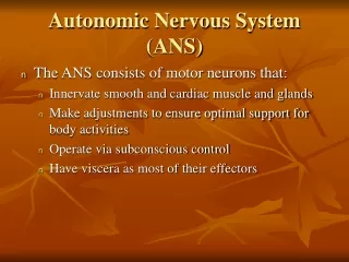
Autonomic Nervous System (ANS). The ANS consists of motor neurons that: Innervate smooth and cardiac muscle and glands Make adjustments to ensure optimal support for body activities Operate via subconscious control Have viscera as most of their effectors. ANS in the Nervous System.

E N D
Autonomic Nervous System (ANS) • The ANS consists of motor neurons that: • Innervate smooth and cardiac muscle and glands • Make adjustments to ensure optimal support for body activities • Operate via subconscious control • Have viscera as most of their effectors
ANS in the Nervous System Figure 14.1
ANS Versus Somatic Nervous System (SNS) • The ANS differs from the SNS in the following three areas • Effectors • Efferent pathways • Target organ responses
Effectors • The effectors of the SNS are skeletal muscles • The effectors of the ANS are cardiac muscle, smooth muscle, and glands
Efferent Pathways • Heavily myelinated axons of the somatic motor neurons extend from the CNS to the effector • Axons of the ANS are a two-neuron chain • The preganglionic (first) neuron has a lightly myelinated axon • The ganglionic (second) neuron extends to an effector organ
Neurotransmitter Effects • All somatic motor neurons release Acetylcholine (ACh), which has an excitatory effect • In the ANS: • Preganglionic fibers release ACh • Postganglionic fibers release norepinephrine or ACh and the effect is either stimulatory or inhibitory • ANS effect on the target organ is dependent upon the neurotransmitter released and the receptor type of the effector
Comparison of Somatic and Autonomic Systems Figure 14.2
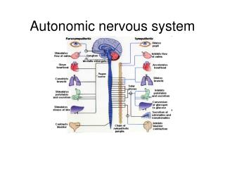
Divisions of the ANS • ANS divisions: sympathetic and parasympathetic • The sympathetic mobilizes the body during extreme situations • The parasympathetic performs maintenance activities and conserves body energy • The two divisions counterbalance each other
Role of the Parasympathetic Division • Concerned with keeping body energy use low • Involves the D activities – digestion, defecation, and diuresis • Its activity is illustrated in a person who relaxes after a meal • Blood pressure, heart rate, and respiratory rates are low • Gastrointestinal tract activity is high • The skin is warm and the pupils are constricted
Role of the Sympathetic Division • The sympathetic division is the “fight-or-flight” system • Involves E activities – exercise, excitement, emergency, and embarrassment • Promotes adjustments during exercise – blood flow to organs is reduced, flow to muscles is increased • Its activity is illustrated by a person who is threatened • Heart rate increases, and breathing is rapid and deep • The skin is cold and sweaty, and the pupils dilate
Sympathetic Outflow • Arises from spinal cord segments T1 through L2 • Sympathetic neurons produce the lateral horns of the spinal cord • Preganglionic fibers pass through the white rami communicantes and synapse in the chain (paravertebral) ganglia • Fibers from T5-L2 form splanchnic nerves and synapse with collateral ganglia • Postganglionic fibers innervate the numerous organs of the body
Sympathetic Trunks and Pathways • The paravertebral ganglia form part of the sympathetic trunk or chain • Typically there are 23 ganglia – 3 cervical, 11 thoracic, 4 lumbar, 4 sacral, and 1 coccygeal
Sympathetic Trunks and Pathways • A preganglionic fiber follows one of three pathways upon entering the paravertebral ganglia • Synapse with the ganglionic neuron within the same ganglion • Ascend or descend the sympathetic chain to synapse in another chain ganglion • Pass through the chain ganglion and emerge without synapsing
Pathways with Synapses in Chain Ganglia • These fibers innervate sweat glands and arrector pili muscles • Rami communicantes are associated only with the sympathetic division
Pathways to the Head • Preganglionic fibers emerge from T1-T4 and synapse in the superior cervical ganglion • These fibers: • Serve the skin and blood vessels of the head • Stimulate dilator muscles of the iris • Inhibit nasal and salivary glands
Pathways to the Thorax • Preganglionic fibers emerge from T1-T6 and synapse in the cervical chain ganglia • These fibers innervate the heart via the cardiac plexus, as well as innervating the thyroid and the skin • Postganglionic fibers emerge from the middle and inferior cervical ganglia and enter nerves C4-C8 • Postganglionic fibers directly serve the heart, aorta, lungs, and esophagus
Pathways with Synapses in Collateral Ganglia • These fibers (T5-L2) leave the sympathetic chain without synapsing • They form thoracic, lumbar, and sacral splanchnic nerves • Their ganglia include the celiac, the superior and inferior mesenterics, and the hypogastric
Pathways to the Abdomen • Sympathetic nerves innervating the abdomen have preganglionic fibers from T5-L2 • They travel through the thoracic splanchnic nerves and synapse at the celiac and superior mesenteric ganglia • Postganglionic fibers serve the stomach, intestines, liver, spleen, and kidneys
Pathways to the Pelvis • Preganglionic fibers originate from T10-L2 • Most travel via the lumbar and sacral splanchnic nerves to the inferior mesenteric and hypogastric ganglia • Postganglionic fibers serve the distal half of the large intestine, the urinary bladder, and the reproductive organs
Pathways with Synapses in the Adrenal Medulla • Fibers of the thoracic splanchnic nerve pass directly to the adrenal medulla • Upon stimulation, medullary cells secrete norepinephrine and epinephrine into the blood
Segmental Sympathetic Supplies Table 14.2
Visceral Reflexes • Visceral reflexes have the same elements as somatic reflexes • They are always polysynaptic pathways • Afferent fibers are found in spinal and autonomic nerves
Visceral Reflexes Figure 14.7
Referred Pain • Pain stimuli arising from the viscera are perceived as somatic in origin • This may be due to the fact that visceral pain afferents travel along the same pathways as somatic pain fibers Figure 14.8
Neurotransmitters and Receptors • Acetylcholine (ACh) and norepinephrine (NE) are the two major neurotransmitters of the ANS • ACh is released by all preganglionic axons and all parasympathetic postganglionic axons • Cholinergic fibers – ACh-releasing fibers • Adrenergic fibers – sympathetic postganglionic axons that release NE • Neurotransmitter effects can be excitatory or inhibitory depending upon the receptor type
Cholinergic Receptors • The two types of receptors that bind ACh are nicotinic and muscarinic • These are named after drugs that bind to them and mimic ACh effects
Nicotinic Receptors • Nicotinic receptors are found on: • Motor end plates (somatic targets) • All ganglionic neurons of both sympathetic and parasympathetic divisions • The hormone-producing cells of the adrenal medulla • The effect of ACh binding to nicotinic receptors is always stimulatory
Muscarinic Receptors • Muscarinic receptors occur on all effector cells stimulated by postganglionic cholinergic fibers • The effect of ACh binding: • Can be either inhibitory or excitatory • Depends on the receptor type of the target organ
Adrenergic Receptors • The two types of adrenergic receptors are alpha and beta • Each type has two or three subclasses (1, 2, 1, 2 , 3) • Effects of NE binding to: • receptors is generally stimulatory • receptors is generally inhibitory • A notable exception – NE binding to receptors of the heart is stimulatory
Effects of Drugs • Atropine – blocks parasympathetic effects • Neostigmine – inhibits acetylcholinesterase and is used to treat myasthenia gravis • Tricyclic antidepressants – prolong the activity of NE on postsynaptic membranes • Over-the-counter drugs for colds, allergies, and nasal congestion – stimulate -adrenergic receptors • Beta-blockers – attach mainly to 1 receptors and reduce heart rate and prevent arrhythmias
Drugs that Influence the ANS Table 14.4
Interactions of the Autonomic Divisions • Most visceral organs are innervated by both sympathetic and parasympathetic fibers • This results in dynamic antagonisms that precisely control visceral activity • Sympathetic fibers increase heart and respiratory rates, and inhibit digestion and elimination • Parasympathetic fibers decrease heart and respiratory rates, and allow for digestion and the discarding of wastes
Sympathetic Tone • The sympathetic division controls blood pressure and keeps the blood vessels in a continual state of partial constriction • This sympathetic tone (vasomotor tone): • Constricts blood vessels and causes blood pressure to rise as needed • Prompts vessels to dilate if blood pressure is to be decreased • Alpha-blocker drugs interfere with vasomotor fibers and are used to treat hypertension
Parasympathetic Tone • Parasympathetic tone: • Slows the heart • Dictates normal activity levels of the digestive and urinary systems • The sympathetic division can override these effects during times of stress • Drugs that block parasympathetic responses increase heart rate and block fecal and urinary retention
Unique Roles of the Sympathetic Division • Regulates many functions not subject to parasympathetic influence • These include the activity of the adrenal medulla, sweat glands, arrector pili muscles, kidneys, and most blood vessels • The sympathetic division controls: • Thermoregulatory responses to heat • Release of renin from the kidneys • Metabolic effects
Thermoregulatory Responses to Heat • Applying heat to the skin causes reflex dilation of blood vessels • Systemic body temperature elevation results in widespread dilation of blood vessels • This dilation brings warm blood to the surface and activates sweat glands to cool the body • When temperature falls, blood vessels constrict and blood is retained in deeper vital organs
Release of Renin from the Kidneys • Sympathetic impulses activate the kidneys to release renin • Renin is an enzyme that promotes increased blood pressure
Metabolic Effects • The sympathetic division promotes metabolic effects that are not reversed by the parasympathetic division • Increases the metabolic rate of body cells • Raises blood glucose levels • Mobilizes fat as a food source • Stimulates the reticular activating system (RAS) of the brain, increasing mental alertness
Localized Versus Diffuse Effects • The parasympathetic division exerts short-lived, highly localized control • The sympathetic division exerts long-lasting, diffuse effects
Effects of Sympathetic Activation • Sympathetic activation is long-lasting because NE: • Is inactivated more slowly than ACh • Is an indirectly acting neurotransmitter, using a second-messenger system • And epinephrine are released into the blood and remain there until destroyed by the liver
Levels of ANS Control • The hypothalamus is the main integration center of ANS activity • Subconscious cerebral input via limbic lobe connections influences hypothalamic function • Other controls come from the cerebral cortex, the reticular formation, and the spinal cord
Levels of ANS Control Figure 14.9
Hypothalamic Control • Centers of the hypothalamus control: • Heart activity and blood pressure • Body temperature, water balance, and endocrine activity • Emotional stages (rage, pleasure) and biological drives (hunger, thirst, sex) • Reactions to fear and the “fight-or-flight” system
Embryonic Development of the ANS • Preganglionic neurons are derived from the embryonic neural tube • ANS structures in the PNS – ganglionic neurons, the adrenal medulla, and all autonomic ganglia – derive from the neural crest • Nerve growth factor (NGF) is a protein secreted by target cells that aids in the development of ANS pathways
Developmental Aspects of the ANS • During youth, ANS impairments are usually due to injury • In old age, ANS efficiency decreases, resulting in constipation, dry eyes, and orthostatic hypotension • Orthostatic hypotension is a form of low blood pressure that occurs when sympathetic vasoconstriction centers respond slowly to positional changes