Download
1 / 40
430 likes | 696 Views
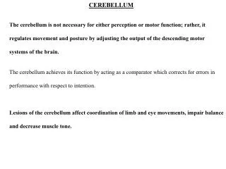
The Cerebellum. Gross anatomy - major divisions - cerebellar peduncles (afferents and efferents) - folia (= gyri) - deep cerebellar nuclei II. Functional anatomy A. Spinocerebellum B. Cerebrocerebellum C. Vestibulocerebellum. III. Regional anatomy

E N D
Gross anatomy - major divisions - cerebellar peduncles (afferents and efferents) - folia (= gyri) - deep cerebellar nuclei II. Functional anatomy A. Spinocerebellum B. Cerebrocerebellum C. Vestibulocerebellum
III. Regional anatomy A. Cytoarchitecture and circuitry of the cerebellar cortex. B. Cerebellar-associated nuclei of the spinal cord + medulla. C. Pontine nuclei. D. Deep cerebellar nuclei. E. Midbrain pathways. F. VLN of thalamus. G. Clinical notes regarding cerebellar lesions.
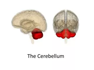
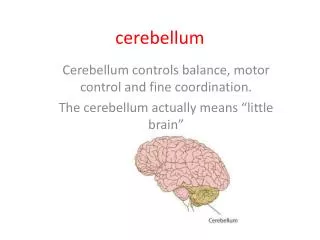
I. Gross Anatomy • Location: dorsal to pons and 4th ventricle and medulla (cut it off to view dorsal surface anatomy of brainstem). - separated from overlying cerebral cortex by tentorium cerebelli (a tough dorsal flap). - 2 symmetrical halves, partly divided by posterior cerebellar incisure (also containing a dorsal flap, the falx cerebri). B. Major anatomical divisions (reflecting functional regions): - vermis - intermediate hemisphere - lateral hemisphere Folds on terminal folia (equivalent to gyri in cerebral cortex) Cortex = organized into groups of folia = 10 lobules and 3 lobes, which are functionally important:
I. Gross Anatomy Anterior: I – V. Posterior: VI – IX. Flocunodular: X. • Cerebellar peduncles (Fig. 13-1B) – All axons travelling to and from the cerebellum course through these. superior: efferents middle: afferents inferior: efferents + afferents D. Deep cerebellar nuclei – key elements of neural circuit (Fig. 13-2B): 4 nuclei (med lat): fastigial globose emboliform dentate
Fig. 13-2B Note: vestibular nuclei also play a role, as we will soon see…
II. Functional Anatomy of the Cerebellum 3 Major functional divisions: A. Spinocerebellum – inputs from sc; controls posture and movement of trunk and limbs. - provides some immediate feedback based on sensory input from the muscles. - comprises the vermis + intermediate hemisphere of both anterior and posterior lobes. - projects through fastigial and interposed nuclei. - has a somatotropic organization. Spinocerebellar tracts: 4 afferent tracts.
II. Functional Anatomy of the Cerebellum: 3 Major Functional Divisions Muscles of body axis Muscles of limbs peripheral
Upper limbs, trunk Fig. 13-6A: Dorsal Spinocerebellar Tract Cuneocerebellar Tract Note from where these tracts originate. Travels in ipsilateral lateral column. Both of these tracts enter cerebellum through the ipsilateral ipsilateral inferior cerebellar peduncle. Lower limbs, trunk Sensory info comes from periphery.
Fig. 13-6B: Descending Pathways: Ventral Spinocerebellar Tract travels after a decussation in the ventral portion of the lateral column and enter cerebellum via the superior cerebellar peduncle. Once in cerebellum, fibres cross again; so, input is ipsilateral! Rostral Spinocerebellar Both relay internal feedback signals reflecting amounts of neural activity in descending pathways. Border of ventral and intermediate zone of sc Lower limbs + trunk
Fig. 13-4: Input-output organization. Inputs: All major inputs feed to dcn, As well as cerebellar cortex. Output = once again through dcn.
Fig. 13-7 Spinocerebellar Output: As noted earlier, the vermis will send efferents through fastigial n. Inferior cerebellar peduncles VL Medial descending pathways and reticulospinal and vestibulospinal 1° motor ctx descends Medial(ventral) Corticospinal tract
Fig. 13-7 The intermediate hemisphere will send efferents through interposed n. Superior cerebellar peduncle Red nucleus (magnocellular) VL Lateral cortico- spinal tract Rubrospinal tract
B. Cerebrocerebellum -participates in the planning of movement -located in the lateral hemisphere -projects to the dentate nucleus This part of the cerebellum is interconnected with the cerebral cortex, rather than receiving its input from the spinal cord. Afferent input – See Fig. 13-8 – from entire contralateral cerebral cortex:
Fig. 13-8 Contralateral cerebral ctx Middle pontine n. Middle cerebellar peduncle Contralateral cerebellar cortex Efferent pathway Dentate n. Red n. (parvocellular) inferior olivary n. (ipsilateral) VL 1° motor ctx and premotor ctx + prefrontal ctx (influences beh and learn)
C. Vestibulocerebellum - functions in maintaining balance and controlling head and eye movements. - input from vestibular labyrinth. - located in floculonodular lobe. - projects to vestivular nuclei (taking the place of the dcn here). Afferent input: • 1° vestibular afferents (note only 1° sensory n. projecting directly to cerebellum). • 2° vestibular neurons in vestibular nuclei. Efferent path: See Fig. 13-9.
Fig. 13-9 Vestibular cerebellar cortex Vestibular nuclei Med longitud Fasciculus (eyes, head) Med and lat VS tracts
III. Regional Anatomy Fig. 13-10 • Cytoarchitecture and circuitry of the cerebellar cortex. - 3 cell layers: molecular, purkinje, granular Folium
5 Cell Types: • Purkinje (inhibitory – GABA) - contacted by climbing fibres - the major output neuron (the only neuron projecting outside the cerebellar cortex). Location: PCL - projects to dcn - projects to vestibular nuclei through inferior cerebellar peduncle (icp). 2. Granule cell (this and remaining 3 are interneurons) - contacted by mossy fibres. - the only excitatory neuron. - puts out just 1 parallel fibre up into molecular layer purkinje dentrites.
5 Cell Types (Cont’d): 3. Stellate Cells – contact Purkinje cell dendrites (inhibitory). - location: outer molecular layer (inhibitory, taurine). 4. Basket Cells – contact Purkinje soma (“basket” around it) – inhibitory. - location: inner molecular layer (inhibitory, GABA). 5. Golgi Cells – contacts granule cell within “glomeruli” (inhibitory) – glial capsule and specificity of connections. - location: granule cell layer (inhibitory, GABA). See Fig. 13-11 for topology of cells.
B. Cerebellar-associated nuclei of the spinal cord and brainstem (medulla). Clarke’s Nucleus and accessory cuneate nucleus relay sensory info to spinocerebellum. • Spinal cord: - Clarke’s n. (Fig. 13-14) courses from C8 to L2 within the medial portion of the intermediate zone. somatic sensory info from lower limbs and trunk - Axons travel in dorsal spinocerebellar tract icp (medulla). - also visible: ventral spinocerebellar tract scp (pons) [originating from spinal border cells].
2. Brainstem: - Accessory cuneate nucleus (Fig. 13-15). - visible in caudal medulla - somatic sensory info from upper limbs and trunk. - trunk served by cuneate fascicle and feeds to icp. Go back and refer to Fig. 13-8: The 1 input is the red nucleus-parvocellular. - Inferior olivary nucleus – origin of all climbing fibres. - Medial and inferior vestibular nuclei (“dcn” for vestibulocerebellum) – receives Purkinje cell axons from flocculonodular lobe vestibulospinal tracts and the medial longitudinal fasciculus (eye muscle control).
C. Pontine nuclei (Fig. 13-16) – These relay input from cerebral cortex cerebrocerebellum. Motor, sensory cortices (1° and 2°) [LAYER 5] pontine nuclei Internal capsule, basis pedunculi axons decussate in pons, later cerebellum via mcp [review: efferent from cerebellum through dentate back to cerebral cortex].
D. Deep cerebellar nuclei - visible in section through caudal pons (Fig. 13-17). - within deep wjite matter underlying the cerebellar cortex. - fastigial n. of pons, medulla (medial descending systems: reticulo, vestibulospinal). - descending projections from all dcn course in the scp.
Fig. 13-17 E. Midbrain pathways 1. Scp and its decussation visible here (caudal midbrain).
Fig. 13-17 2. Interposed n. magnocellular red nucleus rubrospinal tract (note also the corticopontine fibres). 3. Dentate n. parvocellular red nucleus central tegmental tract inferior olivary nucleus. Ascending via the VL to motor cortices and prefrontal cortex (Fig. 13-18).
Fig. 13-18 • VL (thalamic) nucleus. • Cerebellar tract can be followed in Fig. 13-18. • VL is rostral to VPL (for somatic sensory relay). • VL 1° motor cortex and premotor cortex.
G. Clinical notes regarding cerebellar lesions 3 classic signs of cerebellar dysfunction: • Ataxia – inaccuracy (undershoot, overshoot), staggering. • Nystagmus – rhythmic oscillations of the eyes. • Tremors – involuntary oscillations of limbs (“intention tremor”). symptoms are ipsilateral to lesion. [note the many unusual and doubly-crossed pathways] On the next slide, we have some behavioral problems associated with cerebellar lesions