Download
1 / 31
320 likes | 772 Views
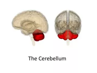
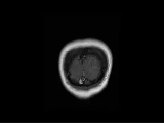
The Cerebellum. Position. Lies above and behind the medulla and pons and occupies posterior cranial fossa Its surface is high convoluted, forming folds or folia, being oriented transversely. Cerebellum. External features.

E N D
Position • Lies above and behind the medulla and pons and occupies posterior cranial fossa • Its surface is high convoluted, forming folds or folia, being oriented transversely Cerebellum
External features Consists of twocerebellarhemisphere united in the midline by the vermis
External features Three peduncles • Inferior cerebellar peduncle -connect with medulla and with spinal cord, contain both afferent and efferent fibers • Middle cerebellar peduncle -connect with pons, contain afferent fibers • Superior cerebellar peduncle -connect with midbrain, contain mostly efferent fibers
External features • Tonsil of cerebellumtwo elevated masses on inferior surface of hemispheral portion just nearby foramen magnum
Lobes • Two deep fissures • Primary fissure • Uvulonodular fissure • Three lobs • Flocculonodular lobe • Anterior • Posterior lobe
Lobes Anterior lobe corpus of cerebellar Primary fissure Posterior lobe Flocculonodular lobe Posterolateral fissure
Internal structures Gray matter • Cerebellar cortex • Cerebellar nuclei • Dentate nucleus • Fastigial nucleus • Emboliform nucleus • Globose nucleus White matter
Internal structures Fastigial nucleus Cerebellar cortex Globose nucleus Dentate nucleus Emboliform nucleus medullary center
Deep Nuclei 1. fastigial nucleus 2. globose nucleus 3. emboliform nucleus 4. dentate nucleus
Three functional divisions • Vestibulocerebellum • Archicerebellum • Flocculonodular lobe • Spinocerebellum • Paleocerebellum • Vermis and intermediate zone • Cerebrocerebellum • Neocerebellum • Lateral zone Intermediate zone Vermis Lateral zone Flocculonodular lobe
Spinocerebellum: Vermis Intermediate hem. Spinocerebellum (Vermis + Intermed. Hem) Cerebrocerebellum: Lateral hem. Control of limbs and trunk Cerebrocerebellum (Lateral hemisphere) Planning of movement+ Vermis Vestibulo-cerebellum (Floculo-nodular lobe) Intermediate hem. Lateral hem. Control of eye & head movements Balance Floculo-nodular lobe Cerebellar divisions IVth vent
Connections and function of cerebellum Vestibulocerebellum • Connections • Afferents: receive input from vestibular nuclei and inner ear. • Efferents: projects to the vestibular nucleus → vestibulospinal → motor neurons of anterior horn • Function: involved in eye movements and maintain balance
Connections and function of cerebellum Spinocerebellum • Connnection • Afferents: receive somatic sensory information via spinocerebellar tracts
Efferents: • Fastigeal reticular and fastigial vestibular pathways: Vermis projects to the fastigial nucleus → vestibular nuclei and reticular formation → vestibulospinal tract and reticulospinal tract → motor neurons of anterior horn • Intermediate zone projects to the interposed nuclei • Globose- emboliform-rubral pathway: Contralateral red nucleus → rubrospinal tract →motor neurons of anterior horn • Function: play an important role in control of muscle tone and coordination of muscle movement on the same side of the body
Connections and function of cerebellum Cerebrocerebellum • Connection • Afferents: receives input from the cerebral cortex via a relay in pontine nuclei(corticopontocerebellar pathway) • Efferents: (dentothalamic pathway): dentate nucleus → contralateral thalamus → primary motor cortex → corticospinal tract → motor neurons of anterior horn • Function: participates in planning movements
summary • Cerebellar efferent fibers: • Globose-emboliform-rubral pathway • Dentothalamic pathway • Fastigial reticular pathway • Fastigial vestibular pathway
summary • Cerebellar afferent fibers: • Afferent fibers from cerebral cortex: corticopontocerebellar pathway. • Afferent fibers from spinal cord: Anterior and posterior spinocerebellar tracts • Afferent fibers from vestibular nerve
Pyramidal Tract and Associated Circuits upper motor neuron UMN Cerebellum BASAL GANGLIA pyramidal tract lower motor neuron UMN
CerebellumFunction Maintenance of Equilibrium - balance, posture, eye movement Coordination of movement of walking and posture maintenance - posture, gait Adjustment of Muscle Tone Motor Learning – Motor Skills
Motor Skill Pablo Casals
CerebellumClinical Syndromes 1-Ataxia: incoordination of movement - decomposition of movement - tremor - past-pointing 2- dysdiadochokinesia 3-Hypotonia, Nystagmus 4- dysarthria
Posture Gait – Ataxia
a b c Cerebellar Ataxia Ataxic gait and position: Left cerebellar tumor a. Sways to the right in standing position b. Steady on the right leg c. Unsteady on the left leg d. ataxic gait d