Download
1 / 9
130 likes | 419 Views
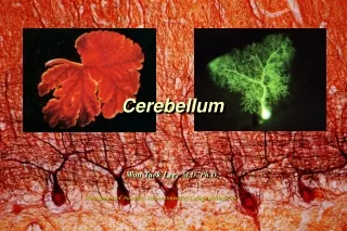
Cerebellum. MIMSA’s Anatom y sessions 2013. cerebellum. Motor part of the brain Coordination of movement Regulation of muscle tone Maintenance of equilibrium Ensures that there is contraction of the proper muscle at the appropriate time and with the correct force. Posterior cranial fossa

E N D
Cerebellum MIMSA’s Anatomy sessions 2013
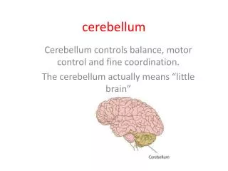
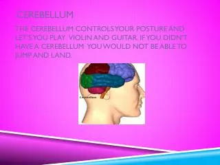
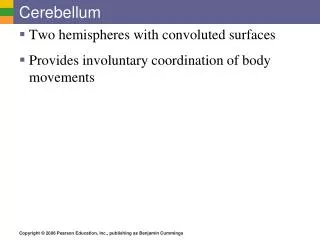
cerebellum • Motor part of the brain • Coordination of movement • Regulation of muscle tone • Maintenance of equilibrium • Ensures that there is contraction of the proper muscle at the appropriate time and with the correct force
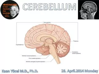
Posterior cranial fossa Separated from the occipital lobes by the tentorium cerebellum Falxcerebelli placed deeply in the posterior cerebellar fissure Fastigium constitutes the roof of the 4th ventricle
Longitudinally: 2 large bilateral hemispheres with vermis between them • Transversally: • Floccolonodular: at the edge of inferior surface; composed of paired irregular-shaped masses – flocula- joined medially by the nodulus (part of the vermis) • Anterior: rostral to the primary fissure. ATTENTION!2nd fissure to develop • posterior lobe: between primary and posterolateral fissures • Posterolateral fissure – between flocculonodular and posterior lobes. 1st fissure to develop
Gray and white matter Gray matter: cortex + 4 types of nuclei in each side White matter: medullary center + paired inferior, middle and superior cerebellar peduncles composed of afferent and efferent nerve fibers which connect the cerebellum with the medulla, pons and midbrain respectively
Cerebellar cortex • Folia cerebelli • 3 layers: • Molecular (stellate +basket) • Ganglionar (purkyne) • Granular (golgi cells + granular cells) • Layers have 5 types of cells: • Stellate • Basket • Purkyne • Golgi • granule
Cerebellar nuclei Transmit all output from the cerebellum Fastigial: close to the midline in contact with the fastigium globulose: 2 or 3 masses in each side Emboliform: oval shape Dentate: most proeminent
White matter • All the afferent (sensory) and efferent (motor) pathways pass through the peduncles. • Inferior cerebellar peduncle • Fibers entering the cerebellum with predominant origin in the inferior olivary complex – olivocerebelar tract; • Middle cerebellar peduncle • Fibers originating in the nuclei pontis / ponto cerebellar tract • Superior cerebellar peduncles • Fibers from globulose, emboliform and dentate nuclei. • Afferent fibers: superior spinocerebellar + rubrocerebellar tracts
Phylogenetical development Archicerebellum – flocculonodular lobe: vestibular nuclei (major connection); function: posture and eye movement Paleocerebellum – superior vermis in the anterior lobe + part of inferior vermis in the posterior lobe; spinal cord (major connection); function: progressive movement Neocerebellum – ceberellar hemispheres + vermis in posterior lobe; cerebral cortex via nclpontis (major connection); funtion: manipulative movement and speech