Download
1 / 53
570 likes | 738 Views
Spinal Cord Tumor. Neurosurgeon Yoon Seung-Hwan, M.D. . 분류 및 호발 부위. 전체 CNS 종양의 15% 경막외 (extradural: ED ) 경막내수외 (intradural extramedullary; IDEM ) 수막내 (intramedullary; IM ). 발생부위별 분류. Secondary carcinoma Primary sarcoma Reticulosis Chordoma Neurinoma Meningioma

E N D
Spinal Cord Tumor Neurosurgeon Yoon Seung-Hwan, M.D.
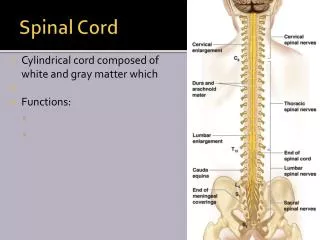
분류 및 호발 부위 • 전체 CNS 종양의 15% • 경막외(extradural: ED) 경막내수외 (intradural extramedullary; IDEM) 수막내 (intramedullary; IM)
발생부위별 분류 Secondary carcinoma Primary sarcoma Reticulosis Chordoma Neurinoma Meningioma Extramedullary Neurinoma Meningioma Epidermoid Intradedullary Ependymoma Astrocytoma Angioma Extradural Intradural
증상 • 임상적인 과정 1) 신경근 시기(root stage) 2) Brown-Sequard stage 3) 척수절단시기(cord transection stage)
운동 기능의 이상 이완성 부전마비 –급속한 진행, 악성 강직성 부전마비 –서서히 진행, 양성
임상 증상과 관련된 요소 1) 부위 – axial location 2) 조직학적 분류 – histological type 3) 침범 정도 – level of involvement
병리조직학적 특징 • Shwannoma Vs Neurofibroma - dorsal nerve root > - 비교적 종양과 신경조직의 분류가 쉽다. - 원형이며 신경에 느슨하게 부착 되어 신경팽대가 없다. ## 10내지 15%에서 dumbbell 형성 (공통)
Neurofibroma neurofibromatosis typeⅠ: von recklinghausens’ disease 과 동반된 심경섬유종의 경우 재발 성향이 강하고 악성화될 가능성이 많다.
Menigioma - arachnoid cap cell에서 origin - calcification - 경막에 비교적 넓게 붙어있다. - 척수의 측면에 위치함 - 척추골 변화 일으키지 않음
Ependymoma - 척수원추에 많이 발생 - 거의 조직학적인 양성 - myxopapillary형이 많다 - 척수와 종양의 분류가 비교적 명확함
Astrocytoma - 조직학적으로 대게 fibrillary 형태 - 소아기 10%, 성인 25%가 악성
Diagnosis • 단순 x-ray erosion of pedicle widening of interpedicular distance enlargement of intervertrbral foramen
요추 천자 Queckenstedt test • 척수 조영술 IM – fish tail appearance IDEM – cup defect ED – paint brush ending
척수혈관 조영술 • 척추 CT • MRI
Schwannoma • Neurilemmoma • 척추강내종양중 가장 많이 발생 – 25% • 주로 IDEM이나 10%는 ED, 1%는 IM 발생 • 30-40대에 많이 발생함
증상 radicular pain이 대게 초기 증상 말기에는 척수기능 상실 증상 --- 하반신 마비, 항문 이상 등
진단 MRI T1w image – iso or hyposignal T2w image – hypersignal Gd – irregular enhancement ring enhacement
Treatment 미세 수술로 완전 적출
Meningioma • 발생부위 thoracic(80%) > 경추(15%) > 요추(5%) • 여자에서 80%이상 • 40-60대에 호발 • 대부분 IDEM 이나 10%정도는 ED • 후측방이나 척수 전방에 위치
증상 신경초종과 증상이 유사 종양이 연수와 경수에 위치 하면 후두부 동통이나 상지 말단부 근위축이 온다.
진단 MRI T1w 과 T2w에서 hypo- or isosignal Gd –균등한 강조영상 을 보이며 tail sign 이 나타난다. • 치료 수술적인 완전 적출
Ependymoma • 척수실질에서 발생하는 종양중 M/C • 증상 국소성 혹은 신경근성 동통 하지 운동 마비나 배뇨장애
진단 MRI 대게 1내지 5개의 척추에 걸쳐 발생 T1w-iso, T2w-hypersignal 균등 enhancement
Treatment 완전 적출 midline myelotomy astrocytoma 보다 good prognosis
Astrocytoma • 발생빈도 및 부위 10세 이하에서 발생하는 척수 종양의 90%이상 차지함 • 증상 운동장애,동통, 감각장애등
진단 MRI T1w - hypo, T2w - iso- Gd –불규칙하고 경계가 명확치 않음 sometimes, syrinx formation
Treatment 75%에서는 양성의 경향을 보이므로 완전 적출시 예후가 좋다 일반적으로 종양의 경계가 불분명하고 10%내외에서 악성인 경우 있음
Metastatic tumor • 발생빈도 20 내지 50%의 발생빈도 대부분 경막외 발생 흉추에 호발 lung > breast > prostate > Kidney
증상 급격한 동통과 급격히 진행되는 신경증상
진단 단순 x-ray - osteolytic or osteoblastic MRI multiple T1w and T2w – low signal Gd 에서 대게 조영증가 소견이 관찰되나 조영증가가 없는 경우도 관찰됨
Treatment steroid decompressive laminectomy 운동마비나 마미증후군 소견
Lipoma • 척수수막류의 형태로 발생하는 경우가 84% 정도임 • 흉추부> 경추부 • 종양이기 보다는 태생기적 유합장애 • 완전 적출시 척수 손상 발생
Hemangioblastoma • 척수 종양의 3% 차지 • 흉수에 60% 이상 • 20%에서 von Hippel-Lindau병과 관련
MRI 척수 팽만 60%에서 낭성 변화 T1w low T2w hypersignal signal void or vascular nodule
Dremoid, Epidermoid, Teratoma • 요천추부 호발 • spida bifida, dermal sinus, 다모증 피부색소침착, 피부혈관종과 관련됨
Chordoma • Notochordal remnant • Sacrococcygeal – 50% Clivus – 35% 척추강 – 15%