Download

1 / 33
420 likes | 737 Views
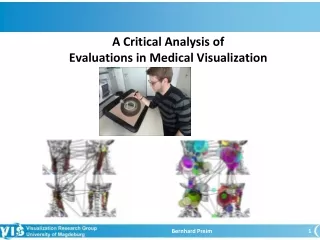
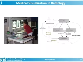
Medical Visualization in Radiology. Radiology technician. Relevant images. Reporting. Outline. Radiology Departments Storage of Medical Image Data Conventional Film-Based Diagnosis Soft-Copy Reading Digital Hanging Protocols 3D Visualizations in Radiology. Radiology Departments.

E N D
Medical Visualization in Radiology Radiology technician Relevant images Reporting Bernhard Preim
Outline • Radiology Departments • Storage of Medical Image Data • Conventional Film-Based Diagnosis • Soft-Copy Reading • Digital Hanging Protocols • 3D Visualizations in Radiology Bernhard Preim
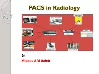
Radiology Departments • Large variety of imaging devices (CT, MR, Computed X-Ray, …) • Large variety of output devices (printer, CRT-monitors with different spatial and grey-value resolution) • Archival of image data in PACS-systems • Huge amounts of data • Imaging devices, output devices, workstations and PACS are interconnected. • Performance problems due to the large amounts of data are frequent and annoying. → Spatial resolution is often reduced to avoid performance problems. Bernhard Preim
Radiology Departments • General radiological workflow Radiology technician Relevant images Reporting Image Courtesy Jens Breitenborn, MeVis Diagnostics Bernhard Preim
Radiology Departments Resulting requirements? • Interoperability of devices from different manufacturers. • Standardized format and description of image data. • Data protection, security and reliable identification of patient and body part, e.g. left leg • „Correct“ or optimal display at different output devices Bernhard Preim
Storage of Medical Image Data • DICOM (Digital Imaging and Communications in Medicine) • Established by NEMA (National Electronics Manufacturer‘s Association) in 1993 • Enables digital communication between imaging devices, information systems and referring physicians (other dept.s of the hospital or external) • Enables exchange of data between devices from different manufacturers • Conformance to the DICOM standard is formally certified. Bernhard Preim
Storage of Medical Image Data Scope of DICOM • 20 parts, some 2200 pages • 26 working groups discuss extensions • Working group 24: Surgical DICOM • Questions, such as: How to represent 3D meshes in DICOM including different representations of an object, application of textures, etc? • Working group 26: Extensions for Digital Pathology • Other working groups: Extensions for representing implant geometries, radiation treatment planning, … Bernhard Preim
Storage of Medical Image Data CT and MRI Data in DICOM • A series of individual DICOM files, each representing a slice. • DICOM files belonging to one dataset are characterized by the same patient-id. • Important tags are included in the legend of a viewer (examples are MeVisLab 2D Viewers). Bernhard Preim
Storage of Medical Image Data Mandatory and optional tags summarized to groups • Patient data: Name, birth date, sex, optional: weight • Image data: study-id, series number, image group • Image presentation: slice distance, pixel spacing, default presentation (window width, window height) • Acquisition parameters: sequence name, special description, reconstruction filter, position and orientation of the patient • Special parameters for a certain modality: e.g. in MR field strength, used coils, echo and repetition time Bernhard Preim
Conventional Film-based Diagnosis Cooperation between radiologist and radiology technician: • Radiologist decides which modality and scanning parameters are used • Radiology technician performs the procedure, operates the device and arranges and prepares the image data for the diagnosis Image Courtesy Sebastian Meyer, MeVis Diagnostics Bernhard Preim
Conventional Film-based Diagnosis Result: • Written report, e.g. using voice recorder • Complex cases: Demonstration for the referring physician and/or interdisciplinary discussion, e.g. tumor board Digital solutions should support these processes! Bernhard Preim
Written report Bernhard Preim
Conventional Film-based Diagnosis • Magnifying glasses are used intensively. • Simultaneous reading of „old“ and „new“ images • Careful and efficient arrangement of image data • Plenty of space for viewing • Films: High spatial and gray level resolution Drawbacks: • Quantitative analysis is heavily restricted. • Diagnostic processes are rather subjective. Bernhard Preim
Conventional Film-based Diagnosis Why is it important? • Radiologists are accustomed to this proces. Digital processes should be somehow „similar“. • Radiologists are extremely efficient in film-based diagnosis. Digital processes must be at least that efficient. • Radiologists cooperate with radiology technicians and focus on the high-level tasks. Thus, the perspective of the radiology technicians is also essential for adopting digital solutions. Bernhard Preim
Soft Copy Reading LCD and CRT monitors: High requirements for CT and MRI viewing, w.r.t. • Display size, • Spatial resolution, • Maximum contrast, • Light intensity, • Quality (very low geometric distortion, very few defect pixels) Careful regular tests including recalibration Bernhard Preim
Soft Copy Reading Special monitors with high spatial resolution (2000x2500 pixels) and high gray value resolution (10 bit) are used for reading X-ray images. • Layout corresponds to the habits in conventional film-based diagnosis • Enables easy comparison between 1-year old and current data. Image Courtesy, MeVis BreastCare, 2002 Bernhard Preim
Soft Copy Reading Image Courtesy, MeVis BreastCare, 2012 Bernhard Preim
Soft Copy Reading Quantitative analysis of gray values in a ROI for assessing the severity of diseases, such as emphysema and fibrotic disease and bone mineral density (osteoporosis) Analysis of cross-sectional areas (severity of a stenosis) Bernhard Preim
Soft Copy Reading • Digital Lightbox (BrainLab) Bernhard Preim
3D Visualizations 3D Visualizations in radiology • Are rarely used at all, since radiologists are trained to infer spatial relations from cross-sectional images. • Support an overview, not an in-depth analyis • Primarily in case of rare anatomic variants, complex fractures. • The radiologist „only“ describes the data. • Are used whenever the referring physician requires it. Preferred viewing modes: • Maximum-intensity projection (often as Movie) • Oblique Multi-planar reformations • Slab rendering (trade-off between 2D and 3D) • Direct volume rendering with predefined TFs Bernhard Preim
3D Visualizations MIP renderings of cerebral MR angiography data. Diagnostic task: search for vascular malformations, e.g. aneurysms Image CourtesyB. Terwey, Bremen Bernhard Preim
3D Visualizations Oblique MPRs and slices Image Courtesy Tobias Boskamp, MeVis Research Bernhard Preim
3D Visualization Orientation cube (size and placement is adjustable). Also sticky figures may be used. Bernhard Preim
3D Visualization Radiation treatment planning (Brainlab) Bernhard Preim
3D Visualization Radiation treatment planning (Brainlab) Bernhard Preim
3D Visualizations Thin Slab-Rendering of CT thorax data. Diagnostic task: search for lung nodules (suspicious of being malignant) Image Courtesy Volker Dicken, MeVis Research Bernhard Preim
3D Visualizations Volume rendering with pre-defined transfer functions (gallery concept) based on high-resolution image data. Image Courtesy, SIEMENS Medical Solutions Bernhard Preim
3D Visualizations Synchronized combinations of 2D and 3D visualizations are useful for treatment planning. Bernhard Preim
3D Visualizations In dentistry, Digital Volume Tomography is employed. Images are also presented in the layout with 4 views. Bernhard Preim
Fused Multimodal Visualizations Clipping planes and lenses in CT data provide a window to underlying MRI data. Also radiation doses are overlaid. Bernhard Preim
Interventional Radiology Bernhard Preim
Summary • The introduction of new techniques in radiology requires careful integration in the infrastructure of radiological departments. • DICOM is essential, also for communicating results. • Analysis of conventional film-based diagnosis is inspiring. • Try to improve on the drawbacks, but also to maintain the positive aspects. • Talk to users, analyze their needs and distinguish between radiologists, radiology technicians and referring physicians. Outlook: • Similar changes occur currently in pathology with much larger microscopy data. Bernhard Preim
Links • Integrating the health care enterprise http://www.ihe.net/ • DICOM http://medical.nema.org/ • DICOMscope - DICOM-Viewer http://dicom.offis.de/dscope.php.de • David Clunie's dicom3toolshttp://www.dclunie.com/dicom3tools.html Bernhard Preim