Download

1 / 17
170 likes | 176 Views
Diagnostic mistakes of radiology visualization of urolithiasis. M. Pasichnyk , S . Pasichnyk, A . Shulyak Lviv National Medical University Lviv, Ukraine. Clinical presentation. Age: 40 years Gender: male Complaints: Acute renal colic .

E N D
Diagnostic mistakes of radiology visualization of urolithiasis M. Pasichnyk , S.Pasichnyk, A. Shulyak Lviv National Medical University Lviv, Ukraine
Clinical presentation • Age: 40 years • Gender: male • Complaints:Acute renal colic. • History of disease: acutely admitted by ambulance to the Rivne Regional Diagnostic Center • History of life: before hospitalization this patient had no complaints. • Occupation: worker of the porcine farm
Results of clinical examination • Pain in right flank, positive Merphy’s sign on the right • Blood cell count: Hb 122 g/L, er 3x10¹²/L, L 6.8x10⁹/L, ESR 9 mm/h • Differentional blood cell count: eosiniphilia (9%). • Urinalysis: normal • Blood biochemistry: normal • Blood coagulation test: normal
Ultrasonography Right kidney: parenchima normal, hydroureteronephrosis to the upper third of ureter (its diameter 9 mm) without any stone Left kidney, liver, spleen, regional lymphatic nodes are normal.
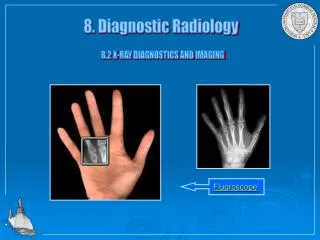
KUB (lumbar region) Please note two shadows suspicious of calculus located at LI and LIII-IV on the right side
KUB (pelvic region) Please note two shadows located in the soft tissues above the coxofemoral joint
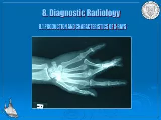
Topogram of the trunk Note the multiple system ossifications in the soft tissues
CT of trunk There are a lot of calcificates in the muscles of back
CT with contrasting Note the hydroureteronephrosis on the right
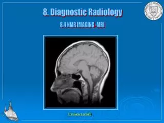
CT of the brain Note foci of calcification in the brain
Conclusions of CT Multiple calcifications of the soft tissues Hydroureteronephrosis to the upper third of the ureter on the right caused by the periureteral calcification (diameter about 7 mm)
Management • This particular patient refused from any treatment • Follow-up of this case was impossible
What is your diagnosis? What additional tests would you prescribe? What management would you suggest?
Our diagnosis • Cystocercosis? • Treatment: specific drugs against cystocercosis according to the consultation of infectionist and ureteral stenting
Similar cases from the literature Typical oval calcifiedcysticercusin muscles of a hip (Nigeria) CT – calcifiedcysticercus located in a brain near the ventriculus, but in that patient not causing other changes (CT with contrasting) (Egypt)
What is cysticercosis ? - Similarly to other parasitic diseases, cysticercosisis caused by the use of contaminated food or water. Sometimes autoinfectionof the patient already infected with a tape worm is noted. Unlike in the brain (or eyes), in the visceral organs cysticercusare surrounded by the fibrous capsule, but remain alive for several years. Holger Pettersson, MD Professor of Radiology University Hospital Lund, Sweden, 1995