Download
1 / 74
800 likes | 1.18k Views
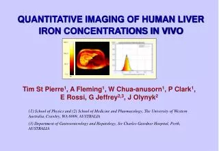
VASCULAR IMAGING OF THE LIVER. Edward G. Grant M. D. Dept. of Radiology University of Southern California Keck School of Medicine. Normal Vascular Anatomy. 3 separate vascular systems PV, HA, HV’s PV 70 - 75% blood / O 2 liver Hepatic artery subordinate

E N D
VASCULAR IMAGING OF THE LIVER Edward G. Grant M. D. Dept. of Radiology University of Southern California Keck School of Medicine
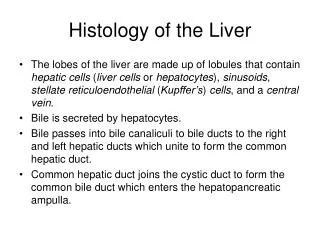
Normal Vascular Anatomy • 3 separate vascular systems • PV, HA, HV’s • PV 70 - 75% blood / O2 liver • Hepatic artery subordinate • Essential in LTX selectively biliary tree • 3 normal hepatic veins IVC • Recognize by anatomy, spectral patterns
Congenital Anomalies &Normal Variants • Normal variants • HA: • Accessory, replaced, separate LHA’s • Commonly arises from SMA, posterior to PV • PV: • Length of extrahepatic PV • HV’s • Inferior/ accessory RHV • Innumerable variations of branching
Congenital Anomalies & Normal Variants • PV aneurysms • HA / PV fistulae • High pressure HA low pressure PV • Symptomatic severe portal HTN • HV / PV fistulae • Low pressure HV low pressure PV • Asymptomatic unless huge encephalopathy • HV / IVC webs ( BCS)
Portal Vein Thrombosis • Color Doppler sonography • Sensitivity = 98%, specificity = 92% • Accuracy = 92% • Negative predictive value = 98% • Good screening technique • Negative Doppler study = no further w/u • Low flow (severe portal HTN) false + • Adjust color sensitivity, use power Doppler Tessler et al. AJR 1991;157:293
Portal Vein Thrombosis & HCC Pozniak et al. Radiology 1991;180:663
Porto-Systemic Shunts • Porto-caval • Portal flow encephalopathy • Selective • Distal spleno-renal (Warren) • Mesocaval • Mesoatrial • Transjugular intrahepatic portosystemic shunt -- TIPS Grant et al.AJR 1990;154:393
TIPS • R / M hepatic vein portal bifurcation • flow, bile leak neointimal hyperplasia stenosis • High failure rate (at HV end) • Routine US surveillance • Q 3 months • Focal flow velocity at stenosis • General velocity in shunt = HV stenosis Dodd et al. AJR 1995;164:1119 Kantermann et al. AJR 1997; 186:467
TIPS / US Contrast Furst et al. AJR 1998;170:1047
The Pulsatile Portal Vein • Abnormal portal vein communications • Porto-systemic shunts • HV / PV fistulae • HA / PV fistulae (more arterialized) • Transmitted cardiac pathology • Right heart failure • Tricuspid regurgitation Duerinckx et al. Radiology 1990; 176:655 Abu-Yousef et al. AJR 1990: 155:785
Budd - Chiari Syndrome • Hepatomegaly, abdominal pain, ascites • Gray scale = vein, color ≠ flow (50%) • Gray scale ≠ vein, color ≠ flow (50%) • Intrahepatic collaterals: “spider web” • Bicolor vein, capsular, portal vein reversed • Blockage ≠ all 3 veins (selective) • PV thrombosis = 20%, IVC blocked = 80% Millener et al. AJR 1993; 161:307
Budd - Chiari Syndrome Bargallo X. et al. AJR 2003; 181:1641