Download
1 / 124
1.35k likes | 1.9k Views
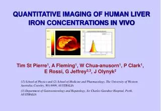
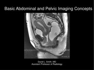
Abdominal Imaging of Liver. Chuan Lu School of Radiology Taishan Medical University. Anatomy Protocols and Normal Ultrasound Findings Pathology. Anatomy of the Liver .

E N D
Abdominal Imaging of Liver Chuan Lu School of Radiology Taishan Medical University
Anatomy • Protocols and Normal Ultrasound Findings • Pathology
Anatomy of the Liver • The Liver occupies all of the right hypochondrium, the greater part of the epigastrium, and left hypochondrium. The ribs cover the greater part of the right lobe .In the epigastric region, the liver extends several centimeters below the xiphoid process. Most of the left lobe of the liver is covered by the rib cage.
Lobes of the Liver • Right lobe: The right lobe of the liver is the largest of the liver’s lobes. It extends the left lobe by a ratio of 6:1. It occupies the right hypochodrium. • Left lobe: The left lobe of the liver lies in the epigastric and left hypochondriac region. • Caudate lobe: The caudate lobe is a small lobe situated on the posterosuperior surface of the left lobe opposite the tenth and eleventh thoracic vertebrae .
Hepatic Nomenclature • Couinaud’s system of hepatic nomenclature provides the anatomic basis for hepatic surgical resection. By using this system , the radiologist may be able to precisely isolate the location of a lesion for the surgical team
Couinaud’s hepatic segments divide the liver into eight segments . The hepatic veins are the longitudinal boundaries . The transverse plane is defined by the right and left portal pedicles .
Hepatic Segmental Anatomy • The caudate lobe (segmentⅠ) is situated posteriorly. • Segment Ⅰincludes the caudate lobe. • Segment Ⅱand Ⅲ includes the left superior and inferior lateral segment. • Segment Ⅳa and Ⅳb includes the medial segment of the left lobe. • SegmentⅤ and Ⅵ are caudal to the transverse plane . • Segments Ⅶ and Ⅷ are cephalad to the transverse plane.
Superior anterior segment (right lobe) Caudate lobe → → Superior lateral segment Superior posterior segment (left lobe) → → Inferior lateral segment → Superior posterior segment (left lobe) → Medial segment → Inferior anterior segment (right lobe)
Ultrasound can allow us to visualize the portal veins, hepatic veins , intrahepatic bile ducts . Anatomy of Liver : Glisson system • The portal veins carry blood from the bowl to the liver, whereas the hepatic veins drain the blood from the liver into the inferior venal cava . The hepatic arteries carry oxygenated blood from the aorta to the liver. The bile ducts transport bile ,manufactured in the liver , to the duodenum.
The portal venous system is a reliable indicator of various ultrasonic tomographic planes throughout the liver.Main portal veinRight main portal veinLeft main portal vein Vascular Supply: Portal veins Intrahepatic Portal Vein Branches • Right anterior superior left median superior • Right anterior inferior left median inferior • Right posterior superior left anterior inferior • Right posterior inferior left lateral superior
Vascular Supply: Hepatic veins The hepatic veins are divided into three components: right,middle,and left. The right hepatic veins is the largest and enters the right lateral
Distinguishing Characteristics of Hepatic and Portal Veins • The best way to distinguish the hepatic from the portal vessels is to trace their points of entry to the liver. The hepatic vessels flow into the inferior vena cava, whereas the splenic veins and superior mesenteric vein join together to form the portal venous system.
Distinguishing Characteristics of Hepatic and Portal Veins • The walls of hepatic veins are thin-walled ,and the walls of portal veins are brightly reflective veins
The hepatic veins are easily differentiated from bile ducts and portal veins . • They are not surrounded by an echogenic wall • They originate close to the diaphragm , and can be traced into the inferior vena cava
Sonographic Evaluation of the Liver • Evaluation of the hepatic structure is one of the most important procedures in sonography for many reasons. The normal , basiclly homogenerous parenchyma of the liver allows imaging of the neighboring anatomic structures in the upper abdomen.
Sonographic Evaluation of The Liver • The system gain should be adjusted to adequately penetrate the entire right lobe of the liver as a smooth ,homogeneous echo-texture pattern • The time gain compensation should be adjusted to balance the far-gain and the near-gain echo signals. • The far time -gain control pods should gradually be increased until the posterior aspect of the liver is well seen.
The appropriate transducer depends on the patient’s body habitus and size • The average adult abdomen usually requires a 3.5MHz
The basic instrumentation should be adjusted in the following parameters : • Time gain compensation • Overall gain • Transducer frequency and type • Depth and focus
Longitudinal Scan Plane • The longitudinal ,or sagittal, scan offers an excellent window to visualize the hepatic structure . With the patient in full inspiration , the transducer may be swept under the costal margin to record the liver parenchyma from the anterior abdominal wall to the diaphragm.
Longitudinal Scan Plane • Scan Ⅰ • Scan Ⅱ • Scan Ⅲ • Scan Ⅳ,Ⅴ,Ⅵ
Longitudinal Scan Plane • Scan Ⅰ The initial scan should be made slightly to the left of the midline to record the left lobe of the liver and the abdominal aorta. The left hepatic and portal veins may be seen as small circular structures in this view.
肝腹主动脉纵切声像图Sagittal image of left lobe of liver, and aorta • The initial scan should be made slightly to the left of the midline to record the left lobe of the liver and the abdominal aorta. The left hepatic and portal veins may be seen as small circular structures in this view.
Sagital image of tip of left lobe of liver The initial scan should be made slightly to the left of the midline to record the left lobe of the liver and the abdominal aorta. The left hepatic and portal veins may be seen as small circular structures in this view.
Sagittal image of left lobe of liver, and aorta The initial scan should be made slightly to the left of the midline to record the left lobe of the liver and the abdominal aorta. The left hepatic and portal veins may be seen as small circular structures in this view. SMA,CA
Longitudinal Scan Plane • Scan Ⅱ • As the sonographer scans at midline or slightly to the right of midline , a larger segment of the left lobe and the inferior vena cava may be seen posteriorly . In this view , it is useful to record the inferior vena cana as it is dilated near the end of inspiration. The left or midline hepatic vein may be imaged as it drain into the inferior vena cava near the level of the diaghram. The area of the portal hepatis is shown anterior to the inferior vena cava as the superior mesenteric vein and splenic vein converge to form the main portal vein. The common bile duct may be seen just anterior to the main portal vein. The head of the pancreas may be seen just inferior to the right lobe of the liver and main portal vein and anterior to the inferior vena cava.
Sagittal image of left lobe of liver, portal vein and inferior vena cava • The left or midline hepatic vein may be imaged as it drain into the inferior vena cava near the level of the diaghram. The area of the portal hepatis is shown anterior to the inferior vena cava
Longitudinal Scan Plane • Scan Ⅲ The next image should be made slightly lateral to this saggital plane to record part of the right portal vein and right lobe of liver . The caudate lobe is often seen in this view.
Gallbladder and Biliary System • Normal size of gallbladder: 7~9cm in length ; 3~4cm in width; Wall thickness : 2~3mm • Normal size of bile ducts : right /left intrahepatic duct just to proximal CHD: 2-3mm ; CBD:≥8mm =dilated
Longitudinal Scan Plane • Scan Ⅳ,Ⅴ,Ⅵ The nest three scans should be made in small increment through the right lobe of the liver . The last scan is usually made to show the right kidney and lateral segment of the right lobe of the liver. The liver texture is compared with the renal parenchyma. The normal liver parenchyma should have a softer , more homogenerous texture than the dense medulla and hypoechoic renal cortex. Liver size may be measured from the tip of the liver to the diaphragm . Generally this measurement is less than 15 cm, with 15 to 20 cm representing the upper limits of normal. Hepatomegaly is present when the liver measurement exceed 20 cm.
肝右肾纵切声像图Sagittal image of liver /right kidney The normal liver parenchyma should have a softer , more homogenerous texture than the dense medulla and hypoechoic renal cortex
The last scan is usually made to show the right kidney and lateral segment of the right lobe of the liver. The liver texture is compared with the renal parenchyma. The normal liver parenchyma should have a softer , more homogenerous texture than the dense medulla and hypoechoic renal cortex.
Transverse Scan Plane • Multiple transverse scans are made across the upper abdomen to record specific areas of the liver. The transducer should be angled in a steep cephalic direction to be as parallel to the diaphragm as possible.
The patient should be in full inspiration to maintain detail of the liver parenchyma , vascular architecture, and ductal structures
Transverse Scan Plane • Scan Ⅰ • Scan Ⅱ • Scan Ⅲ • Scan Ⅳ • Scan Ⅴ,Ⅵ
Transverse Scan Plane • Scan Ⅰ The initial transverse scan is made with the transducer under the costal margin at a steep angel perpenducular to the diaphragm. The patient should be in deep inspiration to adequately record the dome of the liver. The sonographer should identify the inferior vena cava and three hepatic veins as they drain into the cava. This pattern has sometimes been referred to as “reindeer sign” or “Playboy bunny” sign.
The sonographer should identify the inferior vena cava and three hepatic veins as they drain into the cava. This pattern has sometimes been referred to as “reindeer sign” or “Playboy bunny” sign.
Transverse Scan Plane • Scan Ⅱ • The transducer is then directed slightly inferior to the point described in scan Ⅰ to record the left portal vein as it flows into the left lobe of the liver.
Transverse Scan Plane • Scan Ⅲ The porta hepatis is seen as a tubular structure within the central part of the liver. Sometimes the left or right portal vein can be identified . The caudate lobe may be seen just superior to the porta hepatis ; thus , depending on the angel , either the caudate lobe is shown anterior to the inferior vena cava, or as the transducer moves inferior ,the porta hepatis is identified anterior to the inferior vena cava.
Transverse Scan Plane • Scan Ⅳ The fourth scan should show the right portal vein as it divides into the anterior and posterior segments of the right lobe of the liver. The gallbladder may be seen in this scan as an anechoic structure medial to the right lobe and anterior to the right kidney.
肋缘下斜切声像图 The fourth scan should show the right portal vein as it divides into the anterior and posterior segments of the right lobe of the liver. The gallbladder may be seen in this scan as an anechoic structure medial to the right lobe and anterior to the right kidney.
Transverse Scan Plane • Scan Ⅴ,Ⅵ These two scans are made through the lower segment of the right lobe of the liver . The right kidney is the posterior border. Usually intrahepatic vascular structures are not identified in these views
肝脏右叶最大斜径 • 测量标准切面:以肝右静脉和肝中静脉汇入下腔静脉的右肋缘下肝脏斜切面为标准测量切面 • 测量位置:测量点分别置于肝右叶前、后缘之肝包膜处,测量其最大 垂直距离 • 正常参考值:12-14cm
Lateral Decubitus Scan Plane • Left Anterior Oblique • The left anterior oblique scan requires that the patient roll slightly to the left . A 45-degree sponge or pillow may be placed under the right hip to support the patient. This view allows better visualization of the lower right lobe of the liver, usually diaplacing the duodenum and transverse colon to the midline of the abdomen , out of the field of view. Transverse , oblique, or longitudinal scans may be made in this position.
Lateral Decubitus Scan Plane • Measurement of main portal vein • 1.0~1.5cm
Common bile duct • Diameter <0.8cm
Sonographic Evaluation of The Liver • Adequate scanning technique demands that each patient be examined with the following assessment • The size of the liver in the longitudinal plane • The attenuation of the liver parenchyma • Liver texture • The presence of hepatic vascular structures, ligaments ,and fissures