Download

1 / 33
330 likes | 425 Views
COPD. SS Visser, Pulmonology Internal Medicine UP. Chronic Obstructive pulmonary Disease. Two distinct processes are involved, most often in combination. Chronic Bronchitis – dx on history

E N D
COPD SS Visser, Pulmonology Internal Medicine UP
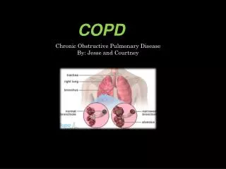
Chronic Obstructive pulmonary Disease • Two distinct processes are involved, most often in combination. • Chronic Bronchitis – dx on history • Emphysema – dx previously on histology, nowadays clinically (good clinical-pathologic-radiologic correlation)
Def: Chronic Bronchitis • Excessive tracheobronchial mucus production sufficient to cause cough with expectoration for most days of at least 3 months of the year for 2 consecutive years. • Classification: • Simple chronic bronchitis • Chronic mucopurulent bronchitis • Chronic bronchitis with obstruction • Chronic bronchitis with obstruction and airway hyperreactivity.
Def: Emphysema • Permanent abnormal distention of air spaces distal to the terminal bronchiole with destruction of alveolar septa (containing alveolar capillaries) and attachments to the bronchial walls. • Classification: • Centriacinar ( centrilobular) emphysema • Panacinar emphysema • Paraseptal emphysema • Senile emphysema
Def: COPD • Chronic obstruction to airflow due to chronic bronchitis and/or emphysema. • Degree of obstruction may be less when the patient is free from respiratory infection and may improve with bronchodilator drugs • Significant obstruction is always present
Epidemiology of COPD • 30% of smokers develop COPD • 20% of adult males have COPD • 15% of COPD patients are severely symptomatic • 4 th leading cause of death (USA) • Mortality rate still rising • prevalence in low birth weight and low socioeconomic status • Tuberculosis in smokers predisposes to COPD
Pathogenesis:Effects of Smoking -1 • Oxidative stress: O2-, OH-,H2O2, HOCl; source of Fe2+ catalizes production of OH- by neutrophils, eosinophils, alveolar macrophages; tar (cigarettes) contains NO and induces iNOStoxic peroxynitrites • Elastin breakdown- activated neutrophils neutrophil elastases and oxidants; -1-AT and metalloproteinase inhibitors (lung defenses) inactivated by smoke • Chemoattractant, upregulation of adhesion molecules neutrophil sequestration in lungs • expression of pro-inflammatory mediators: IL-8, NF-B recruitment of N, B, E and T lymphocytes
Effects of smoking -2 • levels of myeloperoxidase and eosinophilic cationic protein bronchoconstriction • levels of TGF- (transforming growth factor) fibrogenesis • Lipid peroxidation and DNA damage point mutations 0f the p53 gene locus epithelial dysplasia and lung cancer • ciliary function retained secretions; airway resistance vagal-mediated smooth muscle contraction • Hypertrophy and hyperplasia of mucus secreting glands secretions
Pathogenesis-3 • Air pollution exacerbations of CB related to heavy pollution with SO2 and NO2 • Occupation exposure to organic and inorganic dust or noxious gases accelerated decline in lung function • Infection even mild viral respiratory infections ( rhino virus) may be a major factor associated with etiology as well as progression of disease; severe viral pneumonia early in life may lead to COPD • Genetic factors: - -1-antitrypsin deficiency PIZZ, PISZ, PI00 (PI null null), susceptibility to effects of smoking
Pathophysiology • Air trapping- RV and FRC elevated • Hyperinflation –TLC elevated • elastic recoil pressure dynamic collapse of airways during expiration ineffective cough mechanism and pursed lips breathing (emphysema) • compliance (emphysema) • airway resistance • Prolonged forced expiratory time (N=<6 seconds)
Pathology: CB • Hypertrophy of mucus-producing glands in submucosa of large cartilaginous airways • Goblet cell hyperplasia, mucosal and submucosal inflammatory cell infiltrate, oedema, peribronchial fibrosis, intraluminal mucus plugs and increased smooth muscle in small airways • The major site of airflow obstruction is in the small airways and the inflammatory infiltrate consists of neutrophils (in asthma eosinophils)
Pathology : Emphysema • in number and size of alveolar fenestrae eventual destruction of alveolar septa and their attachments to terminal and respiratory bronchioles distention of alveolar spaces • Centriacinar E- respiratory bronchioles (central) affected • Panacinar E- central and peripheral portions of acinus affected • Senile E- alveoli and alveolar ducts enlarge (> 50 Y) • Periacinar/paraseptal E- distention of alveolar spaces adjacent to septal and pleural surfaces
Physical signs of COPD • Ronchi- in early disease present on forced expiration, later present in inspiration and expiration • Prolonged forced expiratory time (> 6 seconds) • Hyperinflation: cardiac dullness, liver dullness displaced downwards, A-P chest diameter, heart and breath sounds, Hoover sign • Inspiratory crepitations (lung bases) • Pursed lips breathing ( dynamic airway collapse) • Use accessory respiratory muscles • Signs of cor pulmonale and PHT
Emphysema = pink puffer Age (Dx) 60 + y Rest dyspnea mild-mod Exer dyspnea severe Cough ± Sputum scanty, mucoid Resp infect less often Resp failure terminal Cor pulmonale terminal Chronic Bronchitis = blue bloater 50 ± y none moderate prominent large volume, purulent often repeatedly common Emphysema:ChronicBronchitis
PHT (rest) 0-mild (exertion) moderate Build Asthenic, cachectic Hematocrit 35-45 Breath pattern use accessory muscles of respiration Sleep pattern Normal XRC Hyperinflation Bullae Mild-moderate severe obese, cyanosed 50-55 do not use accessory muscles of respiration sleep apnea bronchovascular markings Emphysema:Chronic Bronchitis
Blood gas: PaO2± 65 mm Hg PaCO2 35-40 Elastic recoil AW resistance N- Diffusion Cap FEV1 Bronchodilator response Poor 45-60 50-60 Normal N- Better but < 12% and 200ml Emphysema:Chronic Bronchitis
Spirometric classification of COPD severity using post-bronchodilator FeV1 • Stage I (Mild): FeV1/FVC <0.7; FeV1 80% of predicted • Stage II (Moderate): FeV1/FVC <0.7; FeV1 50- <80% of predicted • Stage III (Severe): FeV1/FVC <0.7; FeV1 30-<50% • Stage IV (Very severe): FeV1/FVC <0.7; FeV1 <30% or <50% but chronic respiratory failure is present. (GOLD 2007)
Treatment: Goals of management -1 • Recognition of disease (early Diagnosis and staging) • Smoking cessation (secondary prevention) nicotine replacement and Zyban • Improvement of breathlessness (Rx of airflow obstruction- bronchodilator drugs) 1.Methylxanthines 2.Short and long-acting B2adrenergic agonists ( incidence of pneumonia with ICS and LABA combinations) 3.Short and long-acting Anticholinergics- BD of choice in COPD
Treatment -2 • Respiratory infections –AB when sputum volume and/or purulence (exacerbation of COPD); Influenza and Streptococcus pneumoniae vaccination • Bronchopulmonary drainage and postural drainage (physiotherapy) for patients with CB • Oxygen therapy for patients with hypoxia (PaO2<55 mmHg, SaO2 <88% ) and erythrocytosis (Hematocrit>55) • Pulmonary rehabilitation and education ( improving quality of life)- exercise program and improved nutrition • Prevention and treatment of complications (cor pulmonale) and limitation of disease progression
Treatment -3 • Glucocorticoids –only 10% of COPD patients show subjective benefit and improved lung function (FeV1 increase of 20% or more) on systemic GCs; with COPD exacerbation a course of prednisone 40 mg/d for 2 weeks are usually prescribed • Inhaled GCs may severity of exacerbations and need for hospitalisation. Benefit of 10-14 day trial of 30-40mg prednisone for Stage III COPD patients remains to be proven. • Lung volume reduction surgery • Transplantation
Airway Diseases - COPD • Smoking • Hyperinflation • Airway collapse • Respiratory infection • Bronchospasm • Allergy • Inflammation
Airway Diseases : Asthma • Allergy • Inflammation • Bronchospasm • Hyperinflation • Respiratory infection
AirwayDiseases:Bronchiectasis • Respiratory infection • Hyperinflation • Bronchospasm • Inflammation • Allergy