Download
1 / 19
290 likes | 1.53k Views
Introduction To Corneal Transplantation. Lecture 14 Liana Al-Labadi, O.D. Corneal Transplants. Corneal diseases are the second most prevalent cause of blindness & affect more than 10 million people worldwide according to WHO

E N D
Introduction To Corneal Transplantation • Lecture 14 • Liana Al-Labadi, O.D.
Corneal Transplants • Corneal diseases are the second most prevalent cause of blindness & affect more than 10 million people worldwide according to WHO • The most common treatment for corneal blindness is corneal transplantation with a donor cornea • Corneal Transplant • An operation involving the central part of the cornea • It involves replacing the host cornea with a donor cornea • The operation itself is reasonably straightforward, but the recovery period often takes a long time & optometrists should be able to let their patients know what to expect during and after the surgery
Corneal Transplants • As with cataract surgery, primary care optometrists can develop & refine their clinical skills in comanaging patients who undergo corneal transplant surgery • Understanding & mastering preoperative, intraoperative & postoperative components of each procedure, optometrists can develop solid interprofessional relationships with corneal specialists to improve the patient’soverall treatment & ouctcome
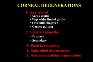
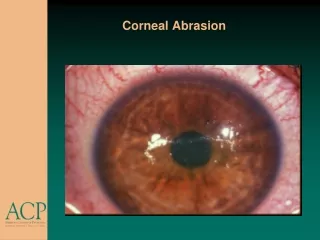
Corneal Layers • Epithelial Layer- 10% of total thickness • Bowman’s membrane- Tough layer • Corneal stroma- Primarily water • Descemet’s membrane- regenerates readily • Endothelial layer- No regeneration
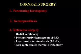
Corneal Transplants • Two major types of corneal transplants • Penetrating keratoplasty (PK)- full thickness • Affected tissue removed full thickness i.e. all corneal layers • Lamellar Keratoplasty- Anterior or posterior • Indicated when a patient has: • Chronic cornea disorder- vision does not meet the patient’s needs • Patient’s vision cannot be corrected with glasses or contact lenses • Patient cannot tolerate his or her correction
Surgical Options • Penetrating Keratoplasty (PK) • Deep Anterior Lamellar Keratoplasty (DALK) • Posterior Lamellar Keratoplasty (PLK) • DLEK • DSEK • DSAEK • DMEK • Keratoprostheses
Corneal Transplants • 1st corneal transplant- performed more than 100 year ago • Corneal transplants at the time involved replacing the entire thickness of the area being transplanted, even when most of the tissue remained healthy • Anterior & posterior lamellar keratoplasty did not become available until the late 90s • Problems with PK surgery • Rejection • High degree of irregular astigmatism • Cataract & glaucoma • PK risks decreased by leaving a portion of the host cornea in place- at least in theory • Pk is indeed a risky procedure • Even more difficult to develop predictable alternatives that provide visual outcomes equivalent to those of PK
Corneal Transplants • % of full-thickness corneal transplant procedures continues to decline • In 2008, over one-third of all corneal grafts were endothelial keratoplasties • % of full-thickness corneal transplant procedures continues to decline • Less risk of rejection & other complications • Recent development in techniques result in visual outcomes more comparable to those of PK • For those active in corneal surgery comanagement, recent developments may leave them confused • Important to analyze the differences between techniques & outcomes of PK & LK to get a better understanding of deep lamellar surgeries
Indications • Indications for corneal transplant may be anatomic or functional • Anatomic indications include: • Visual- opacification, regular refractive error or higher order aberrations (irregular astig) • Reconstructive- thinning or perforation • Therapeutic- edema, dystrophies, degenerations, deposits, intractable infections or painful bullous keratopathy • Cosmetic • Functional Indications • Involve current capabilities, potential capabilities & willingness to risk change • i.e. if an anatomic indication cannot be functionally corrected with medicines or lenses or if the correction cannot be tolerated, a corneal transplant may be warranted
PK Indications • Keratoconus • Keratoglobus • Corneal dystrophies: Macualr, granual & fuch’s dystrophy • Glaucoma • Trauma • Infectious • Corneal scarring or corneal edema • Congenital opacity • Corneal perforation • Bullous keratopathy • Failed Graft
PK Indications • Preoperative consideration • Very important to consider timing of corneal transplant before recommending the surgery • Patients usually experience their best vision 4-8 month postoperatively • Before this time, vision may be worse than before the procedure • Reluctant to recommend surgery on the better eye first or on the fellow eye within six months of the initial procedure • Both eyes rarely require surgery within six months of each other
Outcome • Factors that can complicate the outcome of a corneal transplant include: • Poor eyelid anatomy or function • Severe dry eye • Chemical burns- especially alkaline • Previous radiation treatment • The presence of AC or iris-supported IOL • Elevated IOP • Uveitis • Number of previous grafts • Other surgeries- ex: Radiak Keratotomy • Surgeon’s experience
Outcome • Successful outcome • Significant improvement in the patient’s clinical condition that, in turn, improves his or her overall quality of life • Significant improvement in vision may be two or more smaller lines on an acuity chart • Ability to see with spectacles instead of contact lenses • Restoration of binocularity • Decreased glare • Less pain • Improvement in function & quality of life
Corneal Transplants • Donor corneas • Come from someone who has expressed their wish to donate their corneas to help someone see, after their death • The donor’s cornea will have been thoroughly tested & kept in an Eye Bank for a period, before being sent to the hospital where the operation is to be carried out • Eye Bank is responsible for ensuring the donor cornea is in good condition • Eye Bank performs checks to try and ensure cornea is in good condition • Donor corneas should be free from amy infectious diseases • Must be used within 7 days- placed in sterile moist chamber
The Procedure • Anesthesia • Local block- peribulbar or retrobulbar injection • Used when general health is good • General anesthesia- Used in children • Length of procedure- 1 to 2 hours • During the operation • A circular piece of the host cornea is removed & replaced with a similarly sized piece of the donor’s cornea, which is stitched into place • Other procedures, such as CE, may be done in combination with the corneal graft
The Procedure • Sutures are put into the cornea to hold the new graft in place • They affect cornea’s shape & the way the eye focuses • They are not dissolving sutures & will eventually need to be removed • Two main patterns of sutures are used: • Single running suture- left in place for 1-3 years or until spontaneous breakage occurs • Interrupted sutures- used mostly for corneas with peripheral scarring • can be selectively removed from meridians or areas of vascularization after 2-6 months