Download
1 / 69
700 likes | 724 Views
INTRODUCTION TO HAEMATOPOIETIC STEM CELL TRANSPLANTATION. PROF. SUNDAY OCHENI MB;BS (Ibadan), FMCPath . Consultant Haemato-Oncologist & Haematopoietic Stem Cell Transplantation Physician E-mail: sunday.ocheni@unn.edu.ng. HAEMATOPOIETIC STEM CELL TRANSPLANTATION (HSCT).

E N D
INTRODUCTION TO HAEMATOPOIETIC STEM CELL TRANSPLANTATION PROF. SUNDAY OCHENI MB;BS (Ibadan), FMCPath. Consultant Haemato-Oncologist & Haematopoietic Stem Cell Transplantation Physician E-mail: sunday.ocheni@unn.edu.ng
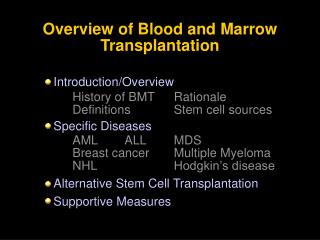
HAEMATOPOIETIC STEM CELL TRANSPLANTATION (HSCT) • HSCT involves the intravenous infusion of normal autologous or allogeneic stem cells collected from bone marrow, peripheral blood, or umbilical cord blood to re-establish hematopoietic function in patients with damaged or defective bone marrow or immune systems or to treat non-haematopoietic disorders
HSCT: Synonyms or related terms • bone marrow transplantation, BMT • peripheral blood progenitor cells, PBPCs • peripheral blood progenitor cell transplantation, PBPCT • stem cell transplantation, SCT • hematopoietic stem cell transplantation, HSCT • autologous proximal stem cell rescue • allogeneic transplant • syngeneic transplant
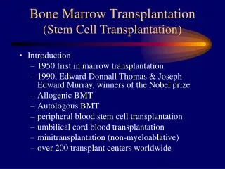
Brief History of HSCT • End of 19th Century: reports of patients given bone marrow orally as a treatment for hematologic disorders and use of orally administered raw or cooked animal spleen • 1939: report of a patient who received 18 mL of intravenous marrow from his brother as a treatment for aplastic anemia • 1950-1962: Report of 203 allo-BMTs, all of which failed
Brief History of HSCT • Late 1960s: discovery of the HLA system • 1968: First successful related allo-SCT. Involved a child with severe combined immunodeficiency treated with stem cells from a sibling donor • 1990: Nobel Prize for Physiology and Medicine shared by Dr. E. Donnall Thomas and Dr. J. Murray for their work in the field of HSCT
TYPES OF HSCT Patient-Donor Relationship • Syngeneic HSCT • Autologous HSCT • Allogeneic HSCT • Haploidentical HSCT Stem-Cell Sources • Bone Marrow Stem Cells • Peripheral Blood Stem Cells • Umbilical Blood Stem Cells
Syngeneic HSCT • From identical twin donors ≈1% of pts • Host and donor are fully matched at both major and minor histocompatibility antigens • Best source of HSCs • No immunological barrier to SCT bc host and donor are genetically indistinguishable • When available, Preferred to auto-HSCT bc there is no risk of tumour contamination of progenitor cells
Syngeneic HSCT2 • Cannot be used for treatment of genetic disorders • Can be used in all circumstances when pt’s progenitor cell population is abnormal on an acquired basis • Low risk of GVHD • Low risk of graft rejection (no immunologic rejection) • Quick posttransplant immune reconnstitution • No risk of tumour cell contamination
Autologous HSCT • Pt’s own HSCs are removed b4 high dose chemo- or radio-therapy and frozen • Cells may be treated (purged) to remove contaminating tumour cells b4 freezing and storage for later use • Purging methods include use of monoclonal antibodies, centrifugation, chemotherapy etc
Autologous HSCT2 • After completing pt’s chemo-or radio-therapy, harvested cells are thawed and returned to the patient • Not applicable to treatment of genetic disorders eg SCD, thalasaemia, storage disorders and congenital immunodeficiencies • Not applicable to situations where most of the progenitor cell population is abnormal on an acquired basis eg MDS, aplastic anaemia and myeloproliferative disorders
Autologous HSCT3 • Lower risk of graft rejection (no immunologic rejection) • No risk of graft-vs-Host disease • No graft-versus leukaemia effect • Quick posttransplant immune reconstruction • No routine posttransplant immunosuppression • Risk of tumour cell contamination
Allogeneic HSCT • Stem cells harvested from another person are infused into the patient in order to reconstitute the haemopoietic and immune systems • Ideally, a (sibling) brother or sister with a similar genetic makeup is used • In the absence of a suitably matched sibling, an unrelated person with a similar genetic makeup may be used
Allogeneic HSCT • Associated with significant morbidity and mortality due to immunological incompatibility bwn patient and donor despite HLA matching • This may manifest as immunodeficiency, GvHD and Graft failure • Paradoxically, there is a Graft vs Leukemia (GvL) effect which probably underlies much of the success of the procedure
Allogeneic HSCT • If pre-transplant immunosuppression is inadequate, Host vs Graft reaction (Graft rejection) may occur • With HLA incompatibility GvHD occurs: the donor stem cells develop an immune reaction and attack some of the patient’s organs. GvHD is the biggest single threat, other than the underlying disease
Haplo-identical HSCT • Donor is father or mother (Half-identical matching) • Very high risk for GvHD
Bone Marrow Transplantation • Source of stem cells is the Bone Marrow • Marrow is usually obtained from the Donor’s posterior iliac crest under anaesthesia • Stem cells are filtered into anticoagulant solutions • Typically, a maximum of 10-15 ml/kg donor body weight (DBW) or 1.5L (whichever is smaller) are collected which gives about 2-5 x 10⁶ CD34+ cells/kg recipient body weight (RBW)
Peripheral Blood Stem Cell Transplantation (PBSCT) • Peripheral Blood Stem Cell Harvest is similar to the process of platelet, red cell, granulocyte or plasma donation using an apheresis machine which removes stem cells from blood by a filtration process • Blood is removed from a vein in one location, filtered, and then returned to a vein in another location. Does not require anaesthesia
Peripheral Blood Stem Cell Transplantation (PBSCT) • Normally, few HSCs circulate in the PB • Administration of haematopoietic growth factors (HGFs) or chemotherapy mobilises many more into d circulation • Healthy doors only receive growth factor, Patients with cancer may receive growth factor alone or chemotherapy plus growth factor.
Peripheral Blood Stem Cell Transplantation (PBSCT) • The most commonly used growth factor is G-CSF NeupogenR • Donors are given 4-5 days of HGFs following which HSCs are collected in one or two apheresis sessions • For auto-SCT, at least 2.5 x 10⁶ CD34+ cells/kg body weight is required
Umbilical Cord Blood HSCT • Cord blood contains a high concentration of HSCs and gives the advantage of immediate availability • Used first time in 1988 to treat a boy with Fanconi anemia; the donor, the boy's newborn sister, was a perfect HLA match for her brother. • Relatively high numbers of hematopoietic stem cells with superior proliferative capacity compared with hematopoietic stem cells from marrow and blood in adults are present in umbilical cord blood collected at the time of delivery
Umbilical Cord Blood HSCT • Owing to the relative immaturity of the immune system in cord samples, stem cells from this source allow the crossing of immunologic barriers that would otherwise be prohibitive. As a result, the degree of tolerable HLA disparity is much greater • However, volume is small and so, HSCs are relatively small. • Limited to paediatric HSCTs • T cells in cord blood are also immature
Umbilical Cord Blood HSCT • Lower incidence of GvHD bc of immature T cells • Slower engraftment compared to other stem cell sources • Another potential problem is the possibility of bacterial contamination in cord blood collections, which could complicate the transplantation procedure • Cord blood is also associated with a low risk of cytomegalovirus positivity; thus, the likelihood of contracting the infection following a cord blood transplantation is primarily related to the recipient's status
If the transplant is successful, the stem cells will migrate into the patient's bone marrow and begin producing new, healthy leukocytes to replace the abnormal cells.
TRANSPLANTATION IMMUNOLOGY • An individual’s immunologic identity is expressed in cell-surface proteins encoded by the cluster of genes known as the Major Histocompatibility Complex (MHC) referred to as Human Leukocyte Antigens in man. • MHC or HLA are critical to the proper function of the immune system • They identify and destroy foreign invaders, while preserving normal healthy tissues.
TRANSPLANTATION IMMUNOLOGY • The success of tissue and organ transplants depends on the donor’s and recipient’s HLA • The HLA proteins are clustered on the short arm of chromosome 6 and have been classified into major categories • HLA-A, HLA-B and HLA-C encode for Class I molecules • The genes in the HLA-DR, HLA-DQ and HLA-DP regions encode for Class II molecules • Class III genes are a heterogenous popn of proteins located bwn Classes I and II
Chromosomes Map HLA
Chromosome 6 -F -G -A -E -C -B - - -TNF -Hsp70 -C2 -Bf -C4 - -DR - -DQ -DM -DP Large number of genes, grouped in different loci, located in a region of the short harm of chromosome 6 POLYGENIC SYSTEM
-F -G -A -E -C -B - - -TNF -Hsp70 -C2 -Bf -C4 - -DR - -DQ -DM -DP 3 classes: • Class I: geni A,B,C,E,F,G,…. • Class II: geni DR,DP,DQ,… • Class III: complement factors… c I III II COMPLEX SYSTEM
TRANSPLANTATION IMMUNOLOGY • Class I antigens are carried on all nucleated cells and platelets. Associated with ß2-microglobulin on the cell surface • Class II proteins are carried on the surface of B-lymphocytes, antigen-presenting cells (macrophages, monocytes, dendritic cells), endothelial cells and activated T-lymphocytes. • Class III genes located bwn HLA I and HLA II and code for : • 2 cytokines- TNF and Lymphotoxin • Complement proteins (C2 and C4 of the classical pathway and factor B of the alternate pathway • A steroid enzyme: 21-hydroxylase
Human Major Histocompatibility Complex - simple map - Cooke & Hill. Nature Rev Genet2001 (www)
Human Leukocyte Antigens • Both Class I and Class II MHC molecules are cell-surface proteins • Their immunologic purpose is to provide a means for the recognition of foreign antigen by the immune system • Foreign antigens (from invading bacteria, viruses etc) are broken down into small peptides by cells. • These small foreign peptides are then brought to the cell-surface “held” by an MHC (HLA) molecule • The HLA molecule is like a cup that holds (and “presents” the foreign antigen to cells of the immune system
Human Leukocyte Antigens • Extracellular (exogenous) foreign antigens (such as bacteria) are “captured” by professional antigen-presenting cells (APCs) , processed within those cells into smaller peptides and then presented on the surface of the APC by a Class II MHC molecule. • Endogenous foreign and other antigens (eg viral products) are degraded within whatever cell harbors the pathogen and then brought to the cell-surface and held by a Class I MHC molecule
Human Leukocyte Antigens • Because CD4 binds to class II MHC molecules, CD4+ helper T cells recognize class II-associated peptides, which are usually derived from ingested proteins. • In contrast, antigens in the cytoplasm are displayed by class I MHC molecules and are recognized by CD8+ cytotoxic T cells, because CD8 binds to class I MHC.
Human Leukocyte Antigens • This segregation of different antigens is key to the specialized functions of CD4+ and CD8+ T cells • The two classes of T cells are designed to combat microbes that are located in different cellular compartments. • Thus HLA class I and class II alloantigens can induce transplant immunity at both humoral (antibody) and cellular (T lymphocyte) immune levels
Classes of MHC Class I: presenters to killer T cells Class II: presenters to helper T cells (APC) Class III: encode for proteins that are secreted, involved in complement system
MHC class I MHC class II Peptide Peptide binding groove Cell Membrane MHC molecules
MHC-encoded -chain of 43kDa 2 1 -chain anchored to the cell membrane 2m Peptide antigen in a groove formed from a pair of a-helicies on a floor of anti-parallel b strands 3 2-microglobulin, 12kDa, non-MHC encoded, non-transmembrane, non covalently bound to -chain, which plays an important role in the structural support of the heavy chain Overall structure of MHC class I molecules HLA-A, -B, -Cw
Chains Structures Structure of MHC class I molecules 1 and 2 domains form two segmented -helicies on eight anti-parallel -strands to form an antigen-binding cleft. Properties of the inner faces of the helicies and floor of the cleft determine which peptides bind to the MHC molecule
MHC-encoded, -chain of 34kDa and a -chain of 29kDa 1 1 a and b chains anchored to the cell membrane 2 2 Peptide antigen in a groove formed from a pair of a-helicies on a floor of anti-parallel b strands Overall structure of MHC class II molecules HLA-DR, DQ, DP No b-2 microglobulin
Peptide a-chain b-chain Cleft is made of both a and b chains MHC class II molecule structure
a-chain a-chain Peptide b-chain Peptide b2-M Cleft geometry MHC class I accommodate peptides of 8-10 amino acids MHC class II accommodate peptides of >13 amino acids
MHC molecules are membrane proteins MHC molecules are able to bind antigenic peptides and to present them to T cytotoxic and T helper limphocytes