Download
1 / 16
221 likes | 682 Views
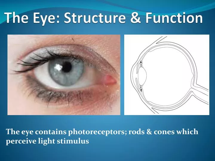
The Eye: Structure & Function. The eye contains photoreceptors; rods & cones which perceive light stimulus. Functions of the eye. the eye allows us to see & interpret the shapes, colors and dimensions of objects in the world by processing the light they reflect

E N D
The Eye: Structure & Function The eye contains photoreceptors; rods & cones which perceive light stimulus
Functions of the eye • the eye allows us to see & interpret the shapes, colors and dimensions of objects in the world by processing the light they reflect • eyes are sense organs that detect and respond to light stimulus giving us sense of sight • they contain photoreceptors; rods & cones located in the retina • rods & cones detects light stimuli & converts light energy to nerve impulses which are transmitted to the brain for interpretation
External Structure of the eye Eye lashes Sclera Pupil Iris Eye lid
Internal Structure of the Eye sclera pupil choroids retina ciliary body / muscles iris lens cornea fovea blind spot optic nerve
Structure Function(s) tough outer layer of the eye which overs and protect eyeball. Sclera Choroid prevents internal reflection of light and nourish retina. Retina contains rods and cones which convert light into nerve impulses. Ciliary Body a ring of muscle controlling the shape and curvature of the lens. Iris controls the pupil size thus controls entry of light. Pupil a hole in the iris that lets light into the back of the eye. Lens accommodation & focusing of light onto the retina. Cornea bends incoming light focusing it on the retina. Fovea a tiny area of densely packed cones for detailed and coloured vision. exit point of the optic nerve cutting through the retina so no rods or cones Blind Spot Optic Nerve carries the impulses from the rods and cones to the visual center of the brain.
The retina: contains rods & cones Axon of the ganglion cell direction of light movement Ganglion cell Bipolar cell Synapse Rod Cone Pigment Sclera
Image formation on the retina • light from an object forms a focused image on the retina • the curved surfaces of the cornea & lens bends the light rays as they pass through them • an image which is smaller than the object is formed upside-down on the retina • the cornea & vitreous humours are mainly responsible for bending of rays of light – refraction while the lens does the final adjustments to focus • lens is elastic & flexible thus it is able to change its shape
Accommodation • the ability of the eye to alter its focus so that clear images of both close and distant objects can be formed on the retina • the shape of the lens can be altered by suspensory ligaments and the ciliary muscles • thickening & thinning of the lens adjusts the focus
Focusing: bending of light rays to fall on the retina Light rays from near object Distance Vision Near Vision Light rays form far object • ciliary muscle relaxes • suspensory ligament stretched by outward pressure of the humours on the sclera • the lens is pulled thin • light from a distant object is focused on the retina – the eye is accommodate i.e. focused for a a distant object • ciliary muscle contracts • suspensory ligament slackens because the ciliary contracts to a smaller circle taking away the tension out of suspensory ligament • the lens is allowed to thicken • light from a near object is focused on the retina – the eye is accommodate i.e. focused for a near object
radial circular CLICK TO CONSTRICT PUPIL DILATED Pupil dilation In dim light, the circular muscles relax& the radial muscles contract, the pupil dilates i.e. widens so that much light is able to reach the retina The pupil is the dark space in the centre of the iris The iris contains circular and radial muscles and their activity can change pupil diameter
radial circular Pupil constriction CLICK TO DILATE PUPIL CONSTRICTED In bright light, the circular muscles contract, & the radial muscles relax, pupil constrictsi.e. narrows so that less light reaches the retina to avoid damage The pupil is the dark space in the centre of the iris The iris contains circular and radial muscles and their activity can change pupil diameter