Download
1 / 75
870 likes | 3.07k Views
Vestibular Examination. ANATOMY & FUNCTION Carmen Casanova Abbott PT, PhD. Lecture Objectives. Discuss vestibular structure as it relates to vestibular function when examining a dizzy patient. Discuss signs and symptoms associated with vestibular disorders

E N D
Vestibular Examination ANATOMY & FUNCTION Carmen Casanova Abbott PT, PhD
Lecture Objectives • Discuss vestibular structure as it relates to vestibular function when examining a dizzy patient. • Discuss signs and symptoms associated with vestibular disorders • Differentiate between peripheral and central vestibular pathology • Discuss components of a physical therapy vestibular examination.
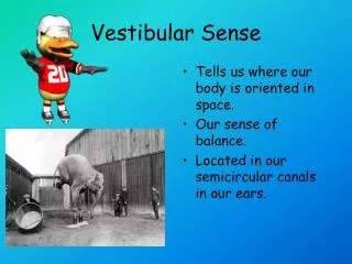
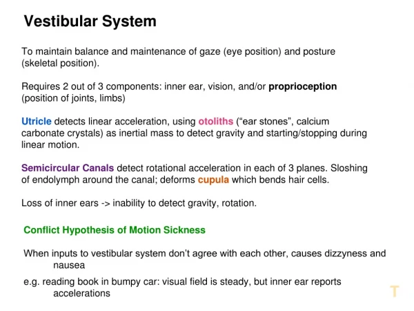
Vestibular System Function • Provides information concerning gravity, rotation and acceleration • Serves as a reference for the somatosensory & visual systems • Contributes to integration of arousal, conscious awareness of the body via connections with vestibular cortex, thalamus and reticular formation • Allows for: • gaze & postural stability • sense of orientation • detection of linear & angular acceleration
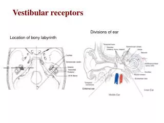
Vestibular Anatomy • Peripheral sensory apparatus • detects & relays information about head angular & linear velocity to central processing system • orients the head with respect to gravity • Central processing system • processes information in conjunction with other sensory inputs for position and movement of head in space • Motor output system • generates compensatory eye movements and compensatory body movements during head & postural adjustments
Peripheral Apparatus • Membranous Labyrinth • Semicircular canals (SSC) • Otolith organs
Semicircular Canals • Angular acceleration • Ampulla contains sensory epithelium
SSC Coplanar Pairing • Spatial arrangement of the 6 SSC cause 3 coplanar pairings • R & L lateral, L anterior and R posterior; l posterior & R anterior; R & L horizontal • Allows for a Push-Pull arrangement of the two sides (e.g., as head turns right, right SSC willincrease firing rate & the left SSC willdecrease firing rate) • Advantages • sensory redundancy • common mode rejection/noise • assist in compensation for sensor overload
Inhibitory Cutoff • Depolarization of the ipsilateral hair cells occurs during angular head movements • Hyperpolarization of contralateral hair cells occurs at the same time • Hair cells are only able to hyperpolarize to what they were at rest = cut off of inhibitory influences from the movement going in the opposite direction even if the ipsilateral hair cells continue to spike higher firing rates
Otoliths • Utricle and saccule • Otolith sensory structures • Maculae • Otolithic membrane • Otoconia • Movement of gel membrane & otoconia cause a shearing action to occur over the hair cells → sensitivity of otoliths
Otolith Function • Respond to: • Linear head motion on acceleration • Static tilt • Two organs respond to respective accelerations or tilts in their respective planes • Saccule has vertical orientation of maculae • Utricle has horizontal orientation of maculae
Hair Cells • 2 types: kinocilium & stereocilia • Sensory structures for the peripheral end organs (maculae and ampula) • Hyperpolarized or depolarized depending upon the direction of deflection of the stereocilia (movement of stereocilia towardsthe kinocilium causes depolarization of the hair cell) • Affect the firing rate of the primary vestibular afferents to the brainstem
Striola of the Macula • Striola serves as a structural landmark • Contains otoconia arranged in narrow trenches, dividing each otolith • Orientation of the hair cells change over the course of the macula • Allows otoliths to have multidirectional sensitivity
Principles of the Vestibular System • Tonic firing rate • Vestibular Ocular Reflex • Push-pull mechanism • Inhibitory cutoff • Velocity storage system
Ascending Pathways • Vestibular nerve • Vestibular nuclei • Cerebellum • Oculomotor complex • CN 3, 4, and 6 • Along with vestibulospinal reflexes coordinate head and eye movements
Relay Centers • Thalamus • Connection with vestibular cortex and reticular formation → arousal and conscious awareness of body; discrimination between self movement vs. that of the environment • Vestibular Cortex • Junction of parietal and insular lobe • Target for afferents along with the cerebellum • Both process vestibular information with somatosensory and visual input
Tonic Firing Rate • Vestibular nerve and vestibular nuclei have a normal resting firing rate (70-100 cycles/sec) • Baseline firing rate present without head movement • Tonic firing is equal in both sides; if not, a sense of motion is felt e.g., vertigo, tilt, impulsion, spinning • Excitation and inhibition of the vestibular system can then occur from stimulation of the hair cells • Spontaneous recovery with light
Vestibular-Ocular Reflex (VOR) • Causes eyes to move in the opposite direction to head movement • Speed of the eye movement equals that of the head movement • Allows objects to remain in focus during head movements
Compensatory Eye Movements • VOR • Optokinetic reflex • Smooth pursuit reflex, saccades, vergence • Neck reflexes • combine to stabilize object on the same area of the retina=visual stability
Vestibular ProcessingGain • Keeps eye still in space while head is moving • Ratio of eye movement to head movement (equals 1)
Vestibular ProcessingVelocity Storage Mechanism • Perseveration of neural firing in the vestibular nerve by the brainstem after stimulation of SSC to increase time constant (10sec.) • SSC respond by producing an exponentially decaying change in neural firing to sustained head movement • Otolith & somatosensory input also drive mechanism
VOR Dysfunction • Direction of gaze will shift with the head movement • Cause degradation of the visual image • In severe cases, visual world will move with each head movement
Oscillopsia • Visual illusion of oscillating movement of stationary objects • Can arise with lesions of peripheral or central vestibular systems • Indicative of diminished VOR gain • motion of images on fovea • diminished visual acuity
Cerebellum • Monitors vestibular performance • Readjusts central vestibular processing of static & dynamic postural activity • Modulates VOR • Provides inhibitory drive of VOR (allowsfor VORc)
Descending Pathways • Provide motor output from the vestibular system to: • Extraocular muscles (part of VOR) • Spinal cord & skeletal muscles (generateantigravity postural activity to cervical, trunk & lower extremity muscles) • Response to changing head position with respect to gravity (righting,equilibrium responses)
Vestibulospinal Reflex (VSR) • Generates compensatory body movement to maintain head and postural stability, thereby preventing falls
Demographics • Vestibular disorders manifested by vertigo are a significant health problem, secondary only to low back pain • NIH study estimates that 40% of the population over the age of 40 will experience a dizziness disorder during their lifetime
Fall Demographics • Falls will be experienced in community dwelling individuals: • 28-35 % over age 65 • 42-49% over age 75 • Greater than 60% will have bilateral vestibular lesion (BVL) in the <65 or >75 years of age
Sedatives Cognitive impairment Palmomental reflex LE disability Foot problems Balance abnormalities Dizziness ↑ dependence on visual cues Fear of falling Orthostatic hypotension (Tideiksaar R 1998) Fall Risk Factors≥ 4 risk factors, 78% chance of falling in an older adult
Aging Changes • Progressive changes begin at age 40 • Decreased number of hair cells • Decreased vestibular nerve fibers • Lead to dizziness and vertigo • Harder to deal with competing visual and somatosensory input
Fear of Falling (FOF) • FOF affects willingness to participate in physical activity & exercise • FOF occurs in an average of 30% of older adults who have not fallen • FOF increases to an average of 60% of older adults who have fallen • FOF is higher among women • Prevalence of FOF is underestimated • Greater FOF associated with lower quality of life in mental health, social & leisure pursuits (Legters, 2002)
Falls Related Self-efficacy • Falls Efficacy Scales (FES) • better for frail • indoor activities • Activities-Specific Balance Confidence Scale (ABC) • higher functioning • indoor & outdoor activities • > discrimination between fearful & nonfearful (Legters, 2002)
Vestibular Pathophysiology • Disorders of tone & or gain (vertigo /movement- induced vertigo) • Vestibular nerve / nuclei give abnormal sensory information • Tone automatically recovers in a few days; does not need visual input • Compensation for reduced gain depends on visual images; takes month to years to complete; high speeds & accelerations may never be complete • Nystagmus usually transient sign of vestibular lesion; movement-induced symptoms can be chronic
Dizzy Patient Presentation: unexplained or new onset of symptoms • Medical referral is indicated • constant vertigo • lateralpulsion • facial asymmetry • speech & or swallowing difficulties • oculomotor dysfunction • vertical nystagmus • severe headaches • recurrent falls • unilateral hearing loss, tinnitus, fullness, ear pain
Vertigo • An asymmetrical firing of the two vestibular systems • Gives an illusion of spinning, movement • Indicative of any one or combination of causes (acute UVH, BPPV, brainstem lesion, vascular hypotension…)
Differentiation Between Peripheral & Central Causes of Vertigo PeripheralCentral Nausea severe moderate Imbalance mild severe Hearing Loss common rare Oscillopsia mild severe Neurologic Symptoms rare common Compensation rapid slow (Furman JM, Whitney SL. 2000)
Peripheral Vestibular Disorders • Vestibular Neuronitis • Labyrinthitis • Meniere’s • Acoustic Neuroma • Fistula • Benign Paroxysmal Positional Vertigo (BPPV)
Central Vestibular Disorders • Vascular • Wallenberg’s Syndrome • Head Injury • Cerebellar Infarct • Postconcussive Syndrome • Demyelinating Disease • Congenital
Degenerative Cerebellar Disease • Signs & symptoms • abnormal ocular pursuit • gradual decline • irregular saccades • gaze end point nystagmus • ataxia
Objective of Clinical Exam • Establish location & severity of lesion (central orperipheral) • Typical examination - history (hearing status) - cranial nerves - vestibular spontaneous nystagmus (imbalance in tone) postural instability (abnormal tone & gain; proprioceptive loss) VOR gain (maintained fixation, dynamic visual acuity) head shaking (compensated UVL; not necessarily PVL) calorics pressure sensitivity (fistula) positional nystagmus (Hallpike-Dix test) hyperventilation (anxiety; acoustic neuroma)