Download
1 / 46
460 likes | 532 Views
Protein Processing in the Endoplasmic Reticulum. Phyllis Hanson Cell Biology Dept., Cancer Res Bldg 4625 phanson22@wustl.edu. Outline. ER morphology Protein folding What happens when protein folding fails ERAD UPR What happens when protein folding is successful

E N D
Protein Processing in the Endoplasmic Reticulum Phyllis Hanson Cell Biology Dept., Cancer Res Bldg 4625 phanson22@wustl.edu
Outline • ER morphology • Protein folding • What happens when protein folding fails • ERAD • UPR • What happens when protein folding is successful • ER exit via COPII vesicles - Retrieval of ER residents from Golgi
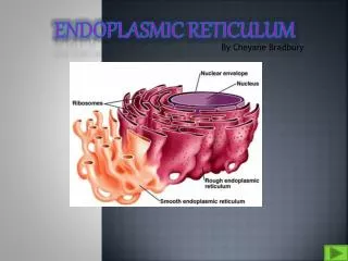
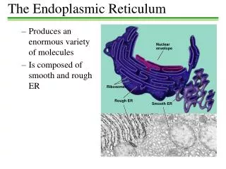
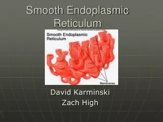
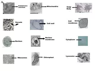
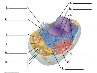
] About 1/3 of cellular protein transits through the ER Subdomains of the ER • Rough ER (mostly ER sheets or cisternae) • Protein translocation • Protein folding and oligomerization • Carbohydrate addition • ER degradation • Smooth ER (mostly ER tubules) • Lipid metabolism • Calcium release • Detoxification • ER exit sites (a.k.a. transitional ER) - export of proteins and lipids into the secretory pathway, marked by COPII coat • ER contact zones - transport of lipids, contact with other organelles • Nuclear envelope • Nuclear pores • Chromatin anchoring
ER subdomains as defined by proximity to other structures Examples from cells expressing fluorescently tagged organelle markers Voeltz lab, UC Boulder
Unfolded protein response ERAD: ER-associated degradation Protein Processing and Quality Control in the Endoplasmic Reticulum Posttranslational modifications Protein folding Unfolded Native Exit from the ER
Protein Modifications and Folding in the ER • Folding challenging in setting of ~400 mg/ml protein concentration • Folding facilitated and monitored by chaperones, both classical (Hsp70/Hsp90) and glycosylation dependent • Folded structure can be stabilized by disulfide bonds, facilitated by protein disulfide isomerases • Final folding can require assembly of multimeric protein complexes
Role of classical chaperones in ER protein folding • ER contains abundant Hsp70 and Hsp90 chaperones • Chaperones help other proteins acquire native conformation, but do not form stable complex • Hsp70s & Hsp90s bind exposed hydrophobic segments • Hsp70 in ER is BiP, interactions with client proteins regulated by ATP hydrolysis and exchange, large variety of cofactors control these • GRP94 is main ER Hsp90, also regulated by ATP status • PPIases–role of prolyl peptidyl cis-trans isomerases
N-linked glycosylation: Asn - X - Ser/Thr Oligosaccharide addition containing a total of 14 sugars Role of glycosylation dependent chaperones in ER folding En bloc addition to protein; subsequent trimming and additions as protein progresses through the secretory pathway; five core residues are retained in all glycoproteins
Increases solubility Fate of newly synthesized glycoproteins in the ER I • Path when nascent protein folds efficiently (green arrows) • Players • OST = oligosaccharyl transferase • GI, GII = glucosidase I and II • Cnx/Crt = Calnexin and Calreticulin, lectin chaperones • ERp57 = oxidoreductase • ERMan1 = ER mannosidase 1 • ERGIC53, ERGL, VIP36 = lectins that facilitate ER exit glucose mannose
Domain structure and interactions of calnexin Model showing interaction of a folding glycoprotein with calnexin and ERp57 Calnexin binds other proteins binds sugar Williams, 2006 J Cell Sci 119:615
Fate of newly synthesized glycoproteins in the ER II • Path when nascent protein goes through folding intermediates (orange arrows) • Players • UGT1 (a.k.a. UGGT) = UDP-glucose–glycoprotein glucosyltransferase, recognizes “nearly native” proteins, acting as conformational sensor • Reglucosylated protein goes through Cnx/Crt cycle for another round • GII removes glucose to try again and pass QC of UGT1 • BiP = hsc70 chaperone that recognizes exposed hydrophobic sequences on misfolded proteins
UDP-glucose glycoprotein glucosyltransferase (UGT1 a.k.a. UGGT or GT) is an ER folding sensor In vitro UGT1 reaction using RNAse as glycoprotein substrate Measure incorporation of [14C] glucose into the oligosaccharide attached to RNAse, compare native vs. denatured RNAse Result: Only denatured RNAse is a substrate. Best substrates in vitro are “nearly folded glycoprotein intermediates” not the native, compact structure or a terminally misfolded protein
Slow Fate of newly synthesized glycoproteins in the ER III • Folding-defective proteins need to be degraded - transported out of the ER for degradation • How do proteins avoid futile cycles? • UGT1 does not recognize fatally misfolded proteins and won’t reglucosylate them for binding to Cnx/Crt • Resident mannosidases will trim mannose residues - protein can no longer be glucosylated and bind to Cnx/Crt • BiP binds hydrophobic regions • Mannosidase trimmed glycans recognized by OS9 associated with ubiquitination machinery • Leads to kinetic competition between folding and degradation of newly synthesized glycoproteins
Protein Processing and Quality Control in the Endoplasmic Reticulum Posttranslational modifications Protein folding Unfolded Native Unfolded protein response Exit from the ER ERAD: ER-associated degradation
ERAD mechanism based on complete reconstitution substrate Stein et al. Cell (2014) 158:1375-138 Baldridge and Rapoport Cell (2016) 166:394-407
What happens when ERAD isn’t enough and misfolded proteins accumulate?
Protein Processing and Quality Control in the Endoplasmic Reticulum Posttranslational modifications Protein folding Unfolded Native Unfolded protein response Exit from the ER ERAD: ER-associated degradation
Unfolded Protein Response (UPR) • Intracellular signal transduction pathways that mediate communication between ER and nucleus • Activated by accumulation of unfolded proteins in the lumen of the ER • First characterized in yeast • Conserved and more complex in animals, with at least three pathways
The UPR in yeast Ire1=inositol-requiring protein-1, ER-localized transmembrane kinase and site specific endoribonuclease Ire1 is maintained in inactive state by binding to BiP. Removal of BiP (by binding to misfolded proteins) leads to Ire1 activation Ire1 activation triggers splicing of intron in mRNA encoding Hac1, a dedicated UPR transcriptional activator Hac1 then binds to UPRE elements to selectively upregulate gene expression of targets that will help alleviate the overload of misfolded proteins
Microarray analysis identified mRNAs for proteins up-regulated by the UPR UPR induced in yeast by treatment with DTT or tunicamycin (Why??) Travers et al. Cell (2000) 150:77-88
Unfolded Protein Response in Metazoans • Three branches • Cells respond to ER stress by: • Reducing the protein load that enters the ER • Transient • Decreased protein synthesis and translocation • Increase ER capacity to handle unfolded proteins • Longer term adaptation • Transcriptional activation of UPR target genes • Cell death • Induced if the first two mechanisms fail to restore homeostasis
UPR also has physiological roles Rutkowski and Hegde, 2010
Misfolded proteins, ER stress, and disease • Cystic fibrosis transmembrane conductance regulator DF508mutation is well studied example (among 100s known) • Protein could be functional as chloride channel at PM, but does not pass ER QC • Ameliorative strategies include use of chemical chaperones, efforts to modulate specific folding factors, and efforts to adjust overall “proteostasis”
UPR as Achilles heel in Multiple Myeloma? • UPR constitutively activated in setting of uncontrolled immunoglobulin secretion • May make cells particularly vulnerable to drugs that interfere with ER stress response, thereby increasing apoptosis • Proteasome inhibitors now in use, p97 inhibitors on the way • Others?
Protein Processing and Quality Control in the Endoplasmic Reticulum Posttranslational modifications Protein folding Unfolded Native Unfolded protein response Exit from the ER ERAD: ER-associated degradation
Minimal COPII machinery Five proteins added to liposomes or in vitro reactions form vesicles: Sar1p, Sec23p, Sec24p, Sec13p, Sec31p
Live cell imaging of VSV-G transport VSV-G ts045 mutant tagged with GFP At 40°C, ts045 VSV-G is retained in the ER due to misfolding Shift to 32°C - traffics to the plasma membrane
Specific amino acid signals mediate selective transport Requirement of two acidic residues in the cytoplasmic tail of VSV-G for efficient export from the ER. Nishimura & Balch, Science 1997
Diacidic motifs are common theme in efficiently secreted proteins VSV-G TM-18aa -YTDIEMNRLGK CFTR TM-212aa-YKDADLYLLD-287aaTM GLUT4 TM-36aa -YLGPDEND LDLR TM-17aa -YQKTTEDEVHICH-20aa CI-M6PR TM-26aa -YSKVSKEEETDENE-127aa E-cadherin TM-95aa -YDSLLVFDYEGSGS-42aa EGFR TM-58aa -YKGLWIPEGEKVKIP-467aa ASGPR H1 MTKEYQDLQHLDNEES-24aaTM NGFR TM-65aa -YSSLPPAKREEVEKLLNG-74aa TfR -19aa -YTRFSLARQVDGDNSHV-26aaTM
Cargo binding sites recognize ER export signals in cytoplasmic domains of cargo Best studied are the diacidic motifs in exiting membrane proteins, but there are others that bind to alternate sites in Sec24 COPII cargo binding site(s) (GAP) (Cargo binding)
What about lumenal cargo? Two possibilities: -bulk flow, with specific retention of ER resident proteins -receptor mediated exit via binding to secreted TM protein
-Measure secretion of soluble model protein derived from Semliki Forest Virus capsid protein -Protein folds rapidly, no need for chaperones -Use pulse-chase analysis to follow newly synthesized protein -First molecule secreted 15 min after synthesis -Rate constant of secretion is 1.2% per minute, corresponding to bulk flow rate of 155 COPII vesicles per second • Soluble proteins are efficiently secreted by bulk flow
And what about large cargo? Malhotra and Erlmann EMBO J 2011 30: 3475
Retrograde traffic from Golgi to ER • Includes receptor-mediated mechanism for retrieving ER resident proteins • HDEL receptor identified in yeast - ERD2; multispanningtransmembrane protein • KDEL receptor in higher euks. • Dilysine motif in C-terminal tail of receptor binds to COPI coat, lumenal domain binds HDEL/KDEL motif in pH dependent manner http://www.ergito.com/lookup.jsp?expt=pelham
Next lectures: Thursday: Mechanism of membrane fusion Secretory pathway organelles and trafficking Tuesday: Endocytic pathways and organelles